History
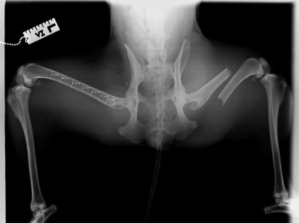
An owned dog was presented to the WVS Malawi clinic with non-weight-bearing lameness of the right hind limb. Her owner explained that on the previous day, the dog had chased after monkeys and returned with non-weight-bearing lameness. The owner had administered paracetamol and brought her to see us the following morning.
Signalment
Age: 3 years
Weight: 22kg
Breed: Border Collie
Sex: Female, entire
Body condition: Ideal (3/5)
Clinical signs
On initial clinical examination, the patient was bright, alert and responsive.
- Mucous membranes were pink
- Body temperature of 38.1°C
- No abnormalities were detected on thoracic auscultation
- Gait examination revealed a 5/5 non-weight-bearing right hind limb lameness.
- Examination of the right hind limb was markedly resented, particularly over the femur. There was also local swelling of the thigh muscles, and instability and crepitus at the level of the femur.
Methadone and meloxicam were given for analgesia and to facilitate further examination.

Differential diagnoses
- Femoral fracture
- Acetabular fracture
- Hip luxation
- Acute patellar luxation
- Cranial cruciate ligament rupture
- Stifle luxation
Based on initial clinical examination, a presumptive diagnosis of femoral fracture was made.
Further investigation
The dog was sedated with medetomidine and induced with a combination of ketamine and diazepam. Radiographs were taken to confirm femoral fracture and rule out other orthopaedic issues.
Diagnosis
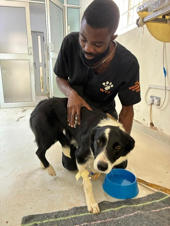
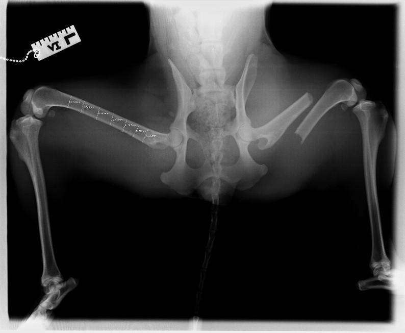
Radiographs revealed a closed, transverse, mid-diaphyseal, severely caudally and proximally displaced right femoral fracture (Figure 2). No other radiographic abnormalities were identified.

Treatment
Decision making for surgical treatment
Several treatment options were considered, which included:
- Lateral plate applied in neutralization or compression mode
- Intramedullary Steinmann pin alone[1], [2]
- Plate with IM pin
- Unilateral (type 1A) external fixator with or without tie-in IM pin and other auxiliary fixation
- Orthogonal plating
- Interlocking nail
- Amputation
Treatment goal and principles
The goal of an open reduction and internal fixation (ORIF) is early comfort and ambulation, complete return of function, and complete bone healing.
For this fracture repair, our goals were:
- Anatomical reduction of the fracture segments
- Stable fixation suitable to the biomechanical and clinical situation
- Preservation of blood supply to the bone fragments and soft tissues
- Early, active pain-free mobilisation of muscles adjacent to the fracture [1], [3]
Surgical treatment
Based on the nature of the fracture and implant availability, lateral plating was preferred. The patient was sedated with medetomidine and methadone, and anaesthesia was induced using ketamine and diazepam. Anaesthesia was maintained by isoflurane in oxygen throughout the procedure. Sciatic and femoral nerve blocks were administered.
During the procedure, the patient received a continuous lidocaine infusion (CRI), IV fluids, and cefoxitin injections every 90 minutes. Cefoxitin was selected in this case based on the clinical circumstances and local drug availability.
Perioperative antimicrobial choice may vary between settings, and should take into account factors such as the procedure, likely pathogens, local resistance patterns, patient factors and available medicines. Vets should use current veterinary antimicrobial stewardship guidance and relevant local or national recommendations to inform their choice. In this case, prophylactic antibiotics were administered given the anticipated long surgical and general anaesthetic time, and use of orthopaedic implants, which are recognised risk factors for postoperative surgical site infection.
Surgical method
A craniolateral approach to the femur was performed to expose the fracture site, and after reduction, the distal and proximal segments were plated with 3.5mm dynamic compression plate (DCP).
1. Lateral approach.
A lateral approach to the femur was used to expose the fracture site [4].
- A craniolateral skin incision was made over the femur, extending from the level of the greater trochanter towards the patella.
- The subcutaneous tissue and superficial fascia were incised in line with the skin incision, and the skin margins were undermined and retracted.
- The fascia lata was incised along the cranial border of the biceps femoris and the incision extended along the required length of the approach.
- The biceps femoris was retracted caudally and the vastus lateralis cranially to expose the femoral shaft.
- The fascial intermuscular septum between these muscles was incised to allow adequate retraction of the vastus lateralis.
- The loose fascial attachments between the vastus intermedius and the cranial surface of the femur were released, allowing the vastus intermedius to be retracted and the fracture site exposed [4].

2. Fracture reduction and bone plating.
Following exposure of the fracture:
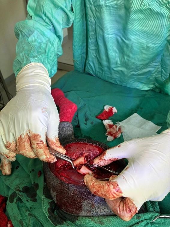
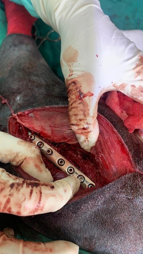
- Bone-holding forceps were used to reduce the fracture and maintain the fragments in anatomical alignment (Figure 3).
- A 3.5 mm dynamic compression plate (DCP) was contoured to match the curvature of the femur and positioned on its lateral surface.
- Four bicortical screws were placed in each fracture fragment using the plate in compression mode [1].
- Pilot holes were drilled using a 2.5 mm drill bit with irrigation. The required screw length was determined by measuring the depth of both cortices before insertion of 3.5 mm non-locking cortical screws.
Plate and screw selection for this case took into account factors including the fracture type and location, the patient's age, activity level and body weight, the size of the bone, and the condition of the surrounding soft tissues [1,5].

3. Closure
- The surgical site was thoroughly flushed before closure.
- The fascia lata was sutured to the cranial border of the biceps femoris.
- The subcutaneous tissue and fascia were closed as a second layer using a continuous suture pattern.
- The skin was closed with a continuous intradermal suture pattern.
Postoperative care
Following the surgery, a Primapore dressing was applied to cover the wound. She received fluid therapy during post-operative recovery. Pain relief was provided as listed below.
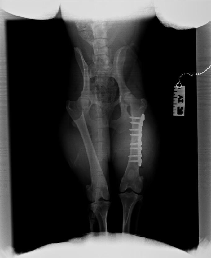
Postoperative radiographs were taken soon after surgery and showed satisfactory fracture reduction, alignment and implant placement (Figure 5).

Analgesia
- Methadone 0.3mg/kg SC q4h was given for two days following surgery.
- Meloxicam 0.1mg/kg PO q24h for a period of three weeks.
- Paracetamol 10mg/kg PO q12h for a period of three weeks.
- Gabapentin 10mg/kg PO q12h for a period of two weeks.
Antibiotics
Cephalexin 15mg/kg PO q12h for five days after surgery
The use of postoperative antibiotics will depend on the individual case and clinical context. Factors such as the degree of tissue trauma or contamination, the nature/length of the surgery, patient-related risks, postoperative care and monitoring, local resistance patterns, and medicine availability may all influence this decision. In uncomplicated cases, postoperative antibiotics may not always be necessary.
An Elizabethan collar was used to prevent her from biting or licking the surgical site. The owner was also instructed to provide strict cage rest for a month and then to walk her on short lead/leash walks for two months.
Prognosis and case outcome
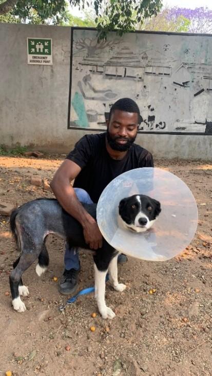
Soon after surgery, the dog was able to toe-touch, and she was weight-bearing a day after surgery (Figure 6).

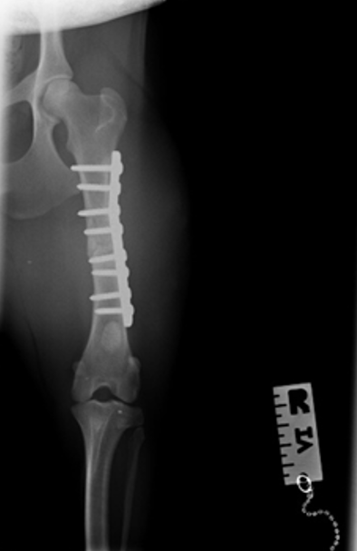
Radiographs were taken three months later, to evaluate how the fracture was healing. The radiographs were used for assessment of alignment, apposition, apparatus and activity. Alignment, apposition and apparatus were all satisfactory. A subtle fracture line remained visible, though fracture healing was considered to be advanced as seen in Figure 7 below.

Discussion
Bone plates are a versatile option for the fixation of many diaphyseal fractures and can provide stable internal fixation. They may be particularly useful in larger dogs, although the choice of fixation method depends on factors including fracture configuration, patient characteristics and implant availability.
The possible risks of the procedure were considered and include:
- Implant failure
- Post-operative infection
- Sciatic nerve damage
- Osteomyelitis
- Implant-associated osteosarcoma, though rare [1]
- Delayed union, non-union or malunion
The favourable outcome in this case was supported by careful preoperative planning, appropriate surgical technique, surgeon expertise, suitable equipment and effective postoperative management.
Key points
- Surgical stabilisation of fractures can support an early return to comfort, limb use and mobility.
- Appropriate surgical planning, and strict adherence to surgical principles and postoperative care guidelines can optimise the outcome.
- Radiographic evaluation is important to assess alignment, apposition, apparatus and activity at the fracture/healing site, 2-3 months following surgery, to help guide continued rehabilitation.
- Implant removal may be considered if complications arise, but routine removal is not usually necessary when implants remain well tolerated.