History
An owned, male entire dog was presented to our clinic by a Mission Rabies vaccination team. He had a three-week history of paraphimosis with penile exposure. According to the history provided, the penis had remained extruded during this period and progressive displacement and retraction of the preputial skin had occurred.
Signalment
Sex : Male, entire
Age : 2 years
Breed : Cross-breed
Weight : 18kg
Body condition : Ideal (3/5)
Clinical examination
On physical examination, the patient was bright, alert and responsive. He had a normal rectal temperature (37.6°C), normal heart rate and sounds (124bpm), and clear lung sounds on auscultation. He was drinking and eating well on his own.
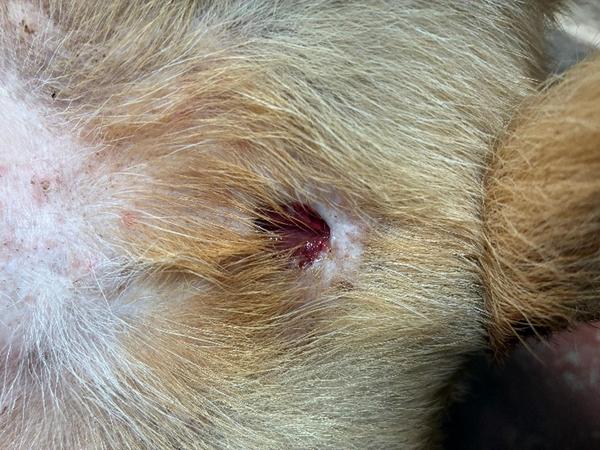
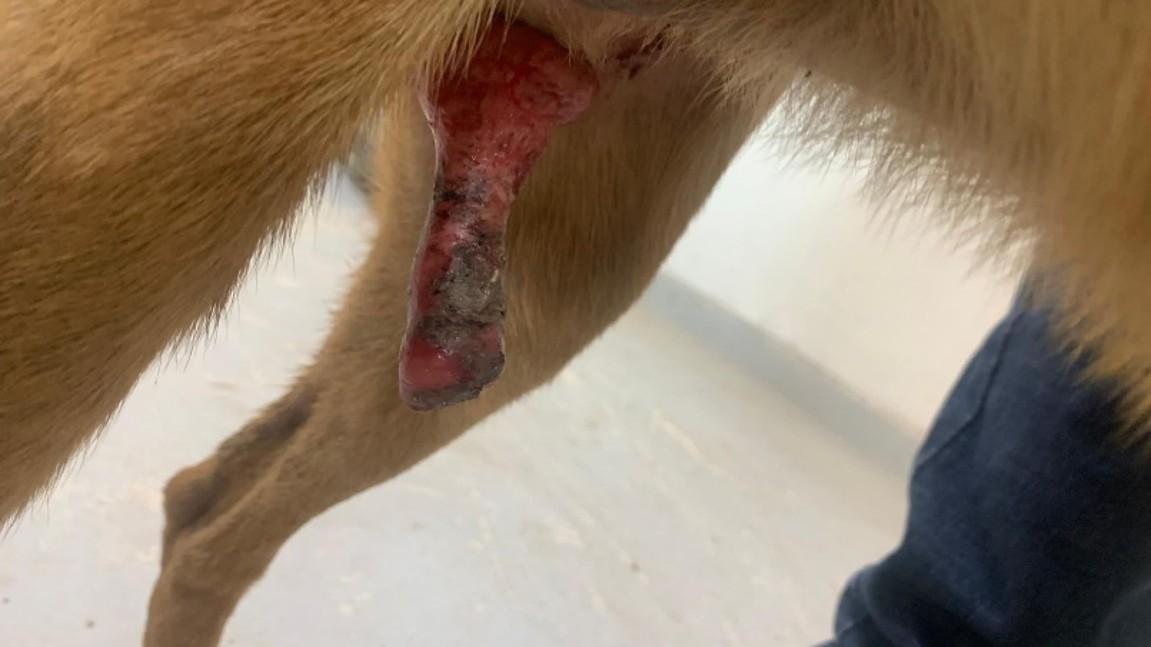
The skin at the preputial orifice was inverted and contaminated with dirt. The patient was ambulatory on admission, with a prolapsed, discoloured, bruised and necrotic penis, as shown in Figure 1. Signs of penile self-trauma were apparent.
Initial treatment
Initial management included application of a commercial lubricant and lavage with a hypertonic sugar solution. Manual reduction of the penis was unsuccessful due to chronic tissue changes and preputial contraction. Broad-spectrum antibiotics (cefalexin 15mg/kg PO q12h) and analgesia (meloxicam 0.1mg/kg PO q24h, and paracetamol 10mg/kg PO q12h) were administered.

Further treatment
Surgical options
A range of surgical techniques may be used in the management of paraphimosis, depending on the underlying cause, chronicity and viability of the penile tissue [1-4]. These include:
- Phallectomy and urethrostomy
- Phallopexy
- Partial phallectomy
- Application of constricting sutures to the preputial orifice
- Preputioplasty
- Cranial prepuce advancement
- Preputial muscle myorrhaphy
Given the chronicity, extensive necrosis, and irreversible tissue damage, complete phallectomy with scrotal urethrostomy was chosen as the surgical treatment.
Anaesthesia and intraoperative management
The patient was sedated with a combination of medetomidine 10µg/kg and methadone 0.3mg/kg IM and induced with a combination of ketamine 2.5mg/kg and diazepam 0.25mg/kg IV. Under general anaesthesia, a rigid urethral catheter was placed retrograde to facilitate identification of the urethra throughout the procedure.
Intraoperatively, the patient received:
- Intravenous fluid therapy
- Continuous rate infusion (CRI) of lidocaine
- Ceftriaxone IV administered every 90 minutes
Surgical procedure
Castration and scrotal ablation
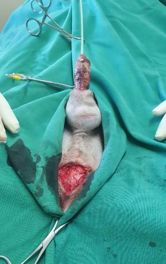
Closed scrotal castration was performed first (Figure 2). An elliptical incision was made at the base of the scrotum, and the entire scrotum was ablated.

Scrotal urethrostomy
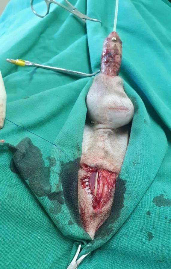
The penile retractor muscle, which runs along the ventral penis and urethral wall, was dissected and retracted to one side to expose the scrotal urethra. The catheter was used as a guide to locate the scrotal urethra, which was opened longitudinally along its ventral midline. A 3cm urethral incision was created (Figure 3). When planning the stoma, allowance should be made for contraction during healing, which may reduce its size considerably. The scrotal region is generally preferred for urethrostomy in dogs because the urethra is relatively superficial and of larger diameter at this level, with less associated haemorrhage than at more proximal sites [4, 5].
On closure, the urethral mucosa and corpus spongiosum were apposed to the cut skin edge with 4-0 polydioxanone monofilament suture material using a continuous suture pattern (Figure 3). A simple interrupted pattern is also possible [1].

Phallectomy
A second elliptical incision was made around the external genitalia, ensuring a minimum 1–2cm margin between the cranial urethrostomy and the caudal aspect of the phallectomy incision, as shown in Figure 4. After identifying and ligating the superficial epigastric vessels with 2-0 polydioxanone monofilament suture, the penis was dissected caudally from the ventral body wall toward the penile bulbs [5]. To ensure haemostasis of the dorsal penile artery, an encircling suture was placed at the amputation site, just proximal to the base of the os penis [4].

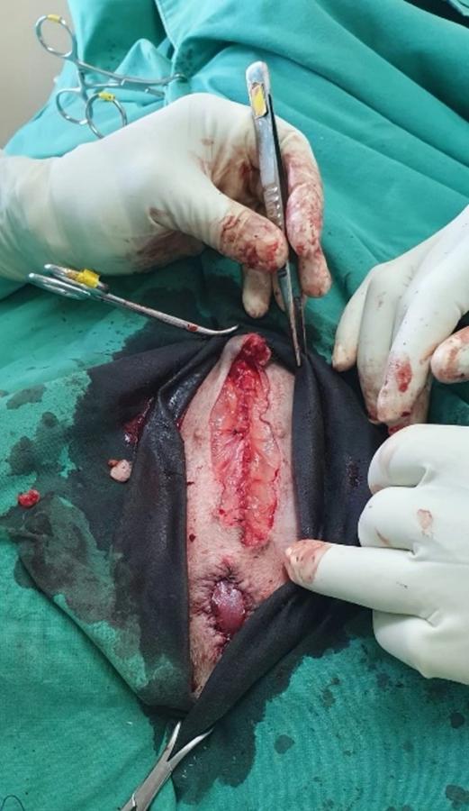
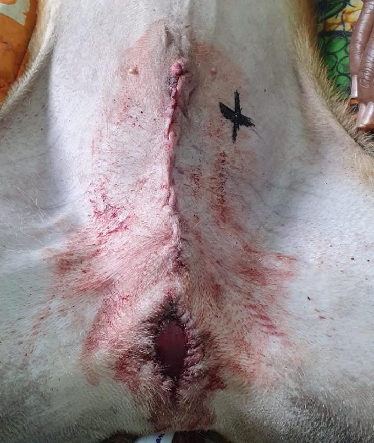
Following amputation, the penile stump was inspected for haemostasis before being allowed to retract caudally into the surrounding connective tissue. The subcutaneous layer was closed in a continuous pattern using a 2-0 polydioxanone absorbable monofilament. Finally, the skin was apposed with a 2-0 polydioxanone using a continuous suture pattern (Figure 5).

Postoperative care
The patient was hospitalised for a week after surgery for close monitoring. Following surgery, he had haemorrhagic discharge from the urethrostomy surgical site for seven days, and the wound was cleaned daily. Haemorrhagic discharge decreased in volume each day. Postoperatively, he received sodium chloride IV fluids until he had fully recovered from the anaesthetic. He also received systemic antibiotics and pain relief.
Analgesia
Postoperative analgesia was tailored to the individual patient, with the following medications administered in this case:
- Methadone 0.3mg/kg SC q4h was given for two days following surgery.
- Meloxicam 0.1mg/kg PO q24h for a period of 3 weeks.
- Paracetamol 10mg/kg PO q12h for a period of 3 weeks.
- Gabapentin 10mg/kg PO q12h for a period of 2 weeks.
Antibiotics
- Ceftriaxone 20mg/kg IV q1.5h intraoperatively
- Cefalexin 15mg/kg PO q12h for 5 days after surgery
The antibiotics used in this case were selected based on the individual clinical circumstances and drug availability. Antimicrobial choice should be made on a case-by-case basis, considering factors such as the suspected infection, patient factors, culture and susceptibility results where available, and local antimicrobial stewardship guidance.
An Elizabethan collar was used to prevent him from biting or licking his surgical site for two weeks.
Prognosis and case outcome
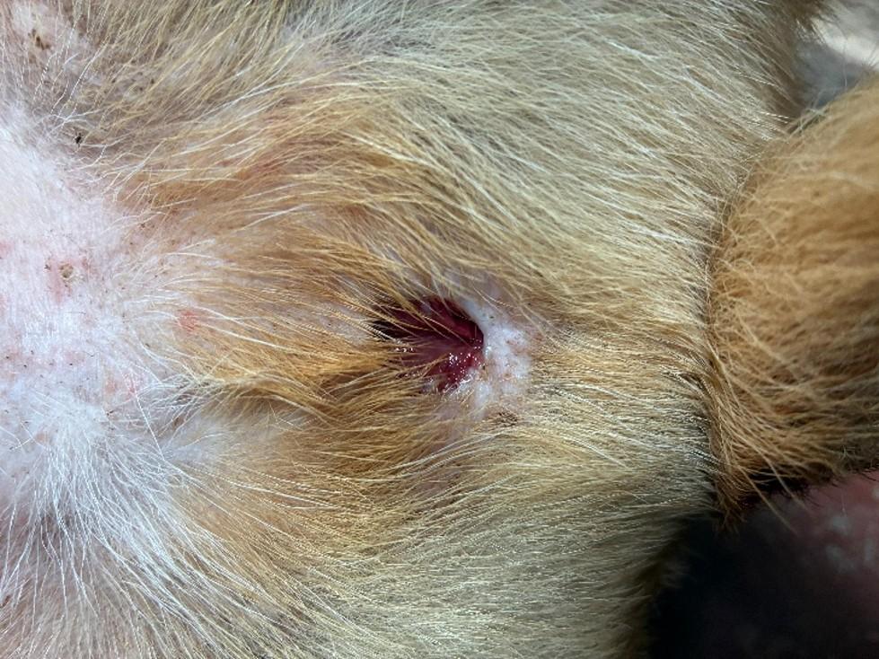
Soon after surgery, the dog was able to urinate without difficulty. The urine was blood-tinged for a few days following surgery. After a week, the haemorrhagic discharge had stopped and grossly normal urine was passed. At a four-month follow-up examination, he was clinically well, urinating without complication, and showed no signs of urinary tract infection (Figure 6).

Discussion
Paraphimosis is a failure to reduce the penis into the preputial skin. Chronic cases can result in vascular compromise, tissue necrosis, and self-trauma.
Strategies to prevent paraphimosis and secondary penile injury include:
- Close supervision of intact males around females in oestrus [1, 3]
- Early veterinary intervention in cases of penile exposure
Early recognition and intervention are essential in cases of paraphimosis to prevent irreversible tissue damage and the need for radical surgery. Indications for phallectomy and scrotal urethrostomy include:
- Severe penile trauma and/or necrosis [1, 5]
- Recurrent urethral obstruction [6]
- Penile neoplasia [1]
Potential complications of the surgery include:
- Haemorrhage
- Surgical site infection
- Wound dehiscence
- Recurrent urinary tract infection
- Urethrostomy stenosis
Conclusion
Although penile trauma is uncommon in male dogs, paraphimosis remains a significant clinical challenge due to the intricate vascular structure of the canine penis and urethra. When irreversible necrosis is present, phallectomy combined with scrotal urethrostomy provides a definitive and effective surgical solution with a good long-term prognosis.
Key points
- The scrotal urethra is considered the optimal site for surgical urethrostomy in dogs based on three key anatomical advantages: diameter, accessibility and haemostasis.
- It is essential to account for postoperative contraction at the urethrostomy site, as the stoma can contract by as much as 50% during the healing process [4].
- Prior to surgery, a retrograde urethral catheter is inserted to enhance visualisation and ensure continuous identification of the urethra throughout the procedure.
- Mild postoperative bloody discharge is normal and should be expected. However, the volume of discharge should steadily decrease each day.