A standard procedure should be followed for evaluating a blood smear to ensure that nothing is missed during the assessment.
What do I need?
To carry out this procedure you will require the following items:
- A stained blood smear (preferably with a coverslip)
- Immersion oil
- A laboratory cell counter (or piece of paper and pen)
- A light microscope with a 10x (dry), 40x (dry) and 100x (oil immersion) objectives
White blood cells
A correctly made blood smear will have the following areas visible (Figure 1):
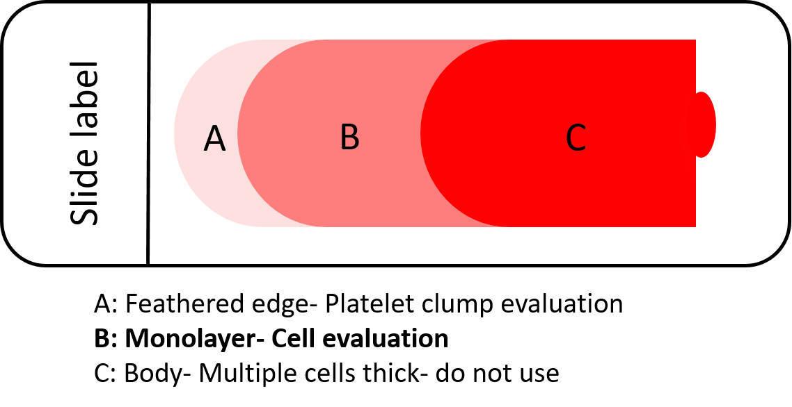
Having a true monolayer enables you to effectively assess the cells. If they are lying on top of each other, this prevents a proper evaluation.
Assessing the featured edge
The purpose of examining the feathered edge is to look for large abnormalities which end up in this area after making the smear. For example, platelets can clump, resulting in an apparent low number in the monolayer, which can lead to a false diagnosis of thrombocytopaenia. In addition, parasites such as microfilaria can be seen here. You should also check that there are not an excessive number of white blood cells (due to poor smear technique), which can result in an inaccurate cell count estimation.
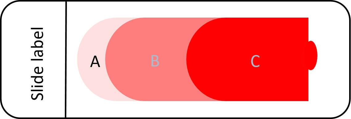
White blood cell counting
The examination and counting of blood cells must be performed in the monolayer. From the feathered edge, move across to this area (Figure 3).
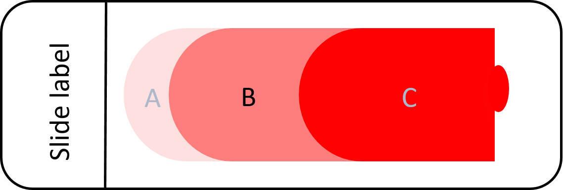
At the junction between the feathered edge and the monolayer you'll notice that the cells form little pools of cells with white spaces between, them giving a 'reticulated' pattern as the cells increase in numbers (Figure 4).

Continue until you are only seeing a continuous monolayer of cells. It is here where you should start counting. If you go too far, you'll see the red cells start to sit on top of each other.
Perform an estimate of absolute WBC numbers. It is important to calculate cell counts (or confirm them, if you already have a readout from a cell analyser). For white blood cells (WBCs), examine the monolayer using a 10x objective. It will provide you with a rough estimate of absolute numbers.
Normal animals will have between 18-50 cells per 10x field in a good quality blood smear. Each WBC corresponds to about 330 cells/μl. Multiply the number of WBC in a 10x field by 330 for an estimation of WBC count.
For dogs, the normal range is between 5.0-14.1 x 10⁹/L SI units (5.0-14.1 x 10³/μl) For cats, the normal range is between 5.2-19.5 x 10⁹/L SI units (5.2-19.5 x 10³/μl)
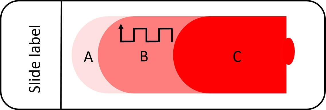
Perform a differential cell count. Cells at both the edges and in the middle of the smear should be included when performing a differential cell count. A good method to include both these areas is to use the battlement meander method of counting (Figure 5).
Count 100 white blood cells and idenfity and record each type (if you can count 200 cells, this will provide you with a more accurate picture). Include nucleated red blood cells and all white blood cells. You will need to use an 100x oil objective for this purpose. If you have an cell counter, this will make it easier; otherwise, record the numbers on a piece of paper.
Once you have finished counting, calculate the percentage of each cell type present.
For example, the number of neutrophils is 55 out of 100. The percentage of neutrophils is therefore 55 / 100 = 0.55 x 100 = 55%
Normally, mature neutrophils will be the predominant cell type, followed by lymphocytes. Only small numbers of eosinophils and monocytes should be present (and rare to absent basophils). The ratio of neutrophils to lymphocytes is approximately 3.5:1 in dogs and 2:1 in cats. Full haematological references ranges for a variety of species can be found here.
Red blood cells
Red blood cells numbers are normally evaluated by assessing packed cell volume (PCV). However, your smear can provide a guide of the RBC density and therefore indicate anaemia. Firstly, visually assess the smear with your eyes against a white background. Does it look pale? Anaemic animals often produce a pale looking smear.
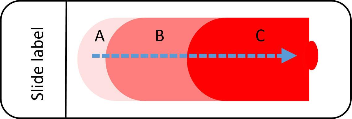
Using the 10x objective, start at the middle of the feathered edge and move back in a straight line through the monolayer counting area and into the body of the slide (Figure 6). In a normal animal, as you move from the monolayer into the body, the RBCs should quickly become piled up on top of each other. However, in an anaemic animal, the cells will not be as densely piled together, and the counting area will be proportionally larger.
RBCs are visually assessed for shape, size, colour and inclusions. Detecting and recognising common abnormalities will be covered in the 'Identifying abnormalities' article.
Platelets
Once you have established that there are no platelet clumps in the feathered edge, platelet numbers can be estimated by counting the total number seen using a 100x objective. It is good practice to repeat this a total of five times using different fields of the monolayer, and then calculate the mean.
Each platelet approximates to 15 x 109/L. For example, if your mean is 12 platelets per 100x objective field, the approximate number of platelets in total is 12 x 15= 180 x 109/L
For dogs, the normal range is between 211-611 x 109/L SI units (5.0-14.1 x 10³/μl) For cats, the normal range is between 300-800 x 109/L SI units (5.2-19.5 x 10³/μl)
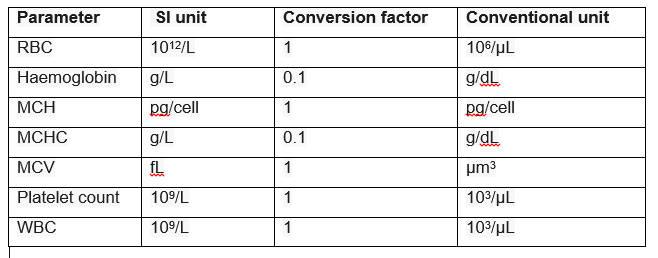
What are conventional and SI units?
Both types of units are used in different countries and it can be confusing when trying to use tables for normal reference ranges. Conventional units are used in the U.S., whereas most countries now use SI units. However, there are conversion factors to help you convert your readings, if you need to.