

History
In general, many of the free-ranging dogs that are operated on in a spay-neuter setting are not formally owned; consequently, there is unlikely to be a detailed history of each dog. In some circumstances, there may be a community caretaker or group of local people that have a degree of interaction with the dogs; and so it is always worth a member of staff asking them if they know of any relevant history, such as recent trauma, behaviour change or the last observed heat. These facts can be useful to know and should be passed on to the operating veterinary surgeon.
Clinical examination
Every animal must be assessed by a clinician prior to surgery.
Clinical examination enables you to assess the risk associated of anaesthesia and surgery for a particular patient. Be consistent in your approach of each body system; this way, you won't miss anything. If there is a wound or obvious problem, continue with the full exam and come back to that at the end. By routinely evaluating normal animals, you will develop a keen eye for spotting anything abnormal.
Free-roaming dogs may not be used to close human contact; they may show fear or aggression and not be easy to handle, or even approachable. Take precautions to keep yourself safe and make sure staff are trained in understanding dog body language.
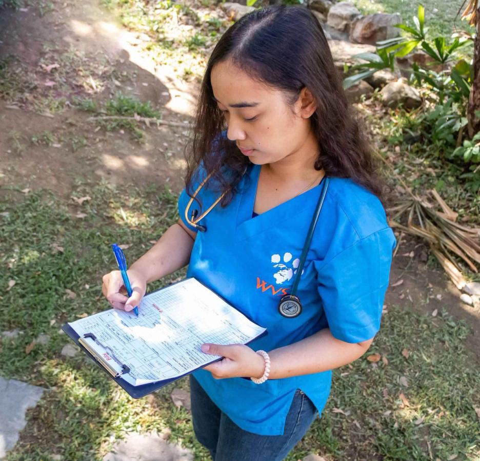
The following should systems be assessed. Remember to wear gloves, especially when assessing the oral cavity in rabies endemic regions. It is often helpful to create a standardised form for assessment which standardises the procedure and ensures that nothing is missed (Figure 1).
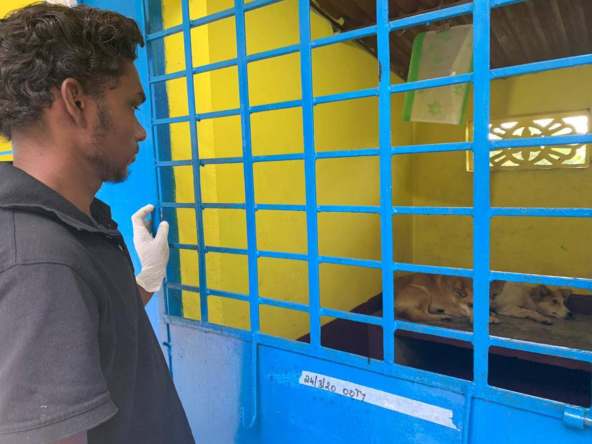
At a distance. Much information can be gained from assessing the patient initially at a distance (Figure 2). Look at the dog's appearance and behaviour; do they look approachable in order to do a 'hands-on' evaluation? Look at posture and gait for signs of abnormalities, such as stiffness, ataxia and lameness. Is there any evidence of behaviours linked to specific diseases, such as rabies or distemper? Any obvious wounds/trauma/tumours/other abnormalities?
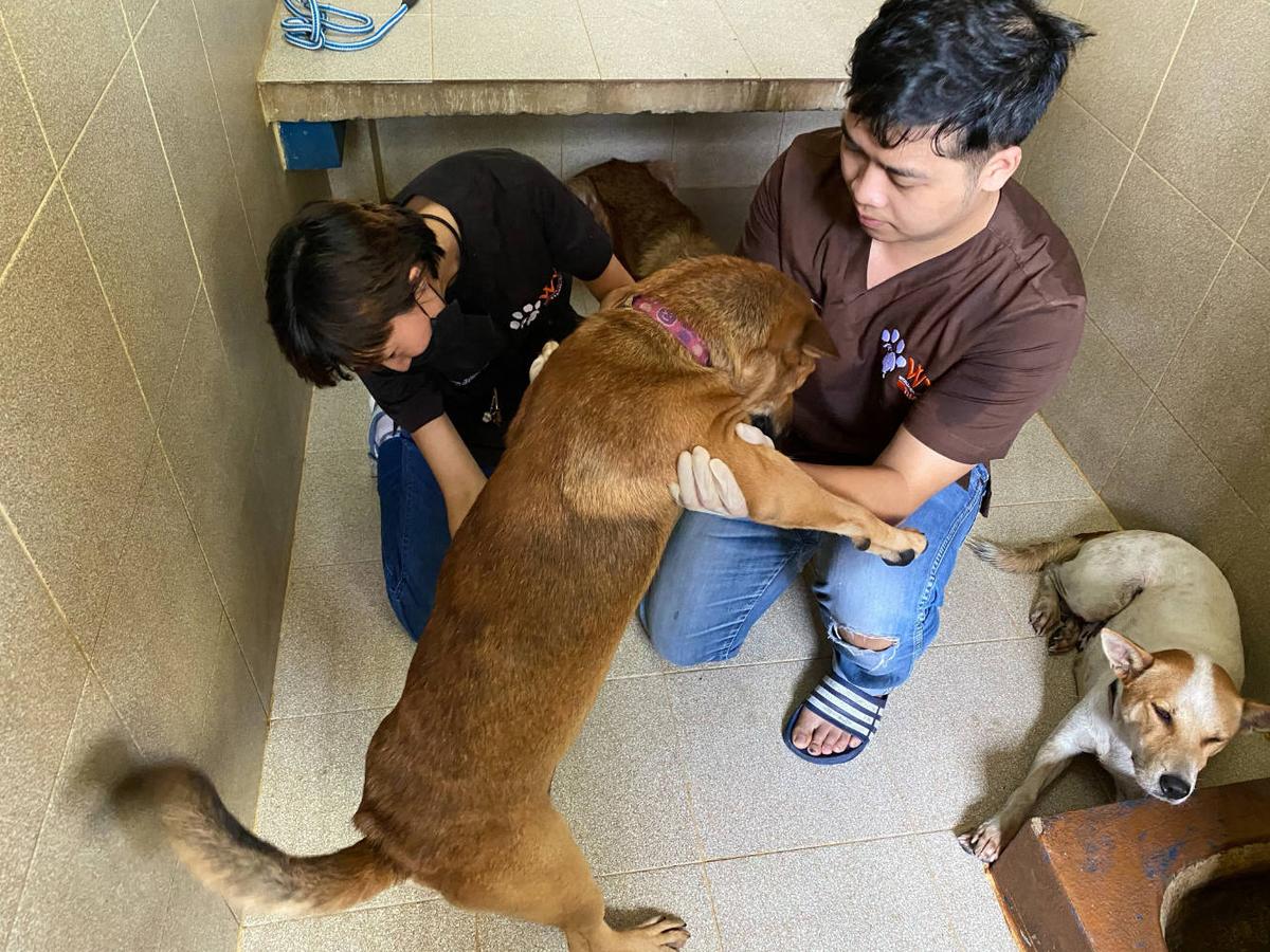
At this point, if the animal's body language suggests that it is safe to approach it, then proceed with the full clinical exam. It is best to start with areas of the body that are easier and less sensitive to touch, to reassure and calm the animal first. For animals whose body language indicate that are not safe to approach, perform this step after sedation (Figure 3).
General condition. Evaluate general condition and assess the body condition score (BCS). There are many different scoring methods for doing this; however, the World Small Animal Veterinary Association's (WSAVA) 'Global Nutrition Panel' has created a 9-point scale has been extensively validated, and is therefore recommended.
Skin. Check for evidence of external parasites such as fleas, lice and ticks. Note abnormalities such as alopecia, pruritus, papules, ulcerations etc. Gently feel all over the body for lumps or wounds. If there is a suggestion of skin disease, skin scrapes and other procedures can be carried out at the end of the exam.
Hydration status. Assess hydration by pinching gently a section of skin on the neck or back (by doing so you are assessing skin turgor). In a well-hydrated animal, this should return rapidly; if an animal is dehydrated, the skin will remained tented for a time. This only becomes apparent once an animal is more than 5% dehydrated. Once you are confident that the animal is calm, you can asses hydration status further by checking the mucous membranes and capillary refill time (CRT) (see oral cavity) and pulse quality. Dogs with tachy mucous membranes can be said to be 5% dehydrated, progressing to skin tent and slightly prolonged CRT (6-8% dehydration), and dry mucous membranes, sunken eyes, bounding pulses, loss of skin turgor and alteration of consciousness (10 - 12% dehydration) Anaesthetising animals that are dehydrated poses a greater risk of low blood pressure and renal damage, particularly with the use of NSAIDs. For mild dehydration your intra-operative fluid therapy plan should be adjusted, but more marked dehydration should be corrected before anaesthesia is performed.
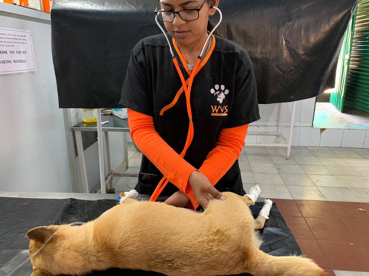
Thorax. Observe and palpate the thorax for symmetry, and the presence of masses. Calculate the heart rate and, at the same time, palpate the pulse (e.g. inguinal) to ensure there are no inconsistencies between the two. Listen for abnormalities such as an arrhythmia, murmur or muffled heart sounds (Figure 4). To assess the respiratory system, listen the the lungs on both sides; move the stethoscope around on the chest to cover the whole lung field and evaluate the respiratory rate, depth, effort. Listen in at least four locations.
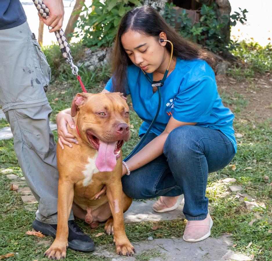
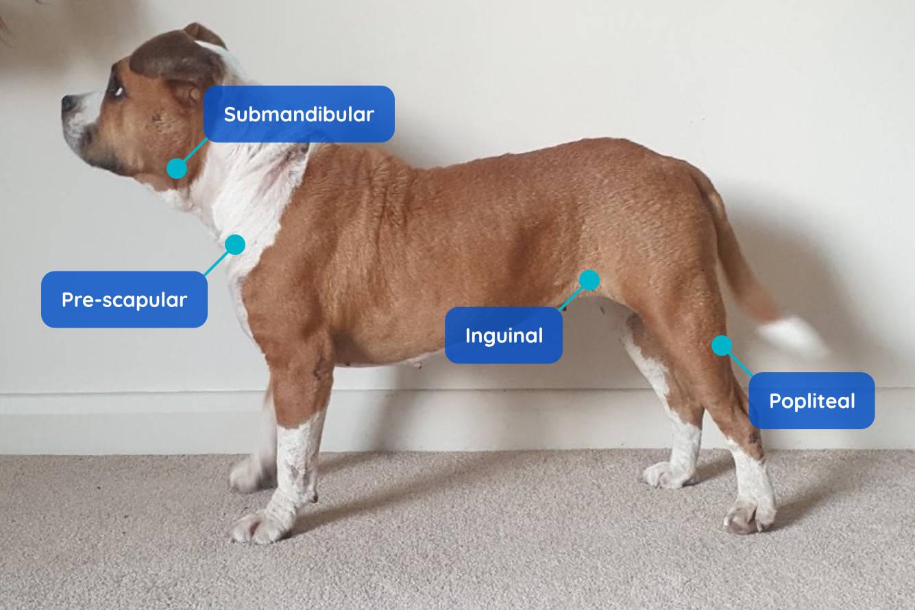
Peripheral Lymph Nodes. Locate and palpate the sub-mandibular, prescapular, popliteal and inguinal lymph nodes to check for enlargement or pain (Figure 5). The axillary (in the axilla or 'arm-pit' region is not normally palpable in the healthy animal, but the area should still be palpated.
Eyes. Evaluate the colour of the sclera, look for the presence of an elevated third eyelid, the colour of the conjunctiva, abnormal discharges, blepharospasm (increased blinking), epiphora (increased tear production), pupil size and symmetry, and assess vision.
Ears. Both ears should be odourless, and clean. Excessive brown or other coloured discharges are abnormal. Assess for the presence of pain, inflammation, external parasites, foreign bodies etc. Is there an ear notch from previous neutering surgery? Failure to spot an ear notch in a female dog will put her through an unnecessary surgical procedure.
Oral cavity. Be cautious when assessing the mouth and always wear gloves, especially in rabies endemic settings. Mucous membrane (mm) colour and capillary refill time (CRT) will provide useful information about the current, cardiovascular and hydration status. Also, briefly assess teeth, gums, jaw tone tumours, halitosis (bad breath that may indicate dental or other disease), and salivation.
Mucous membrane colour: Oral gingiva (gums) should be a pink colour and moist in a healthy animal. Pale mucous membranes can indicate anaemia or poor perfusion (this can be due to conditions such as haemorrhage or circulatory collapse). Reddened or congested membranes (cyanosis) can be due to conditions such as congestion, fever or sepsis. Yellow or icteric membranes can result from a blockage in bile flow, liver disease, or due to increased red blood cell destruction.
Capillary refill time: Apply pressure to the patient's gums to blanch them. When your finger is removed, the colour should return in 1-2 seconds. If this is prolonged, it indicates poor perfusion due to hypovolaemia or to dehydration.
Neck. Gently palpate the trachea; applying slight pressure can result in a cough, which may indicate an infection or an anatomical issue. You may consider postponing surgery to avoid exposing other animals in the clinic to kennel cough.
Abdomen. Gently palpate for the presence of fluid, gas, masses, excessive faecal material or the presence of foetus(es), indicating pregnancy. Note any pain or guarding of the abdomen.
Limbs. Palpate each limb and joint, noting any abnormalities, such as pain, heat swelling, masses, crepitus or injuries.
Genital Organs. Confirm the gender and if male, check to ensure both testes are present; for females, see if a previous spay scar is visible or palpable (Figure 6). Check for abnormalities such as a transmissible venereal tumour (TVT) or other tumours, as well as trauma, abnormal discharges etc. Assess the vulva for abnormal discharge that could indicate a pyometra.
Temperature. If an animal looks unwell, take its rectal temperature. However, in healthy dogs, this step can be omitted.
Normal parameters for dogs
Normal clinical parameters for dogs are summarised below:
| Parameter | Adult | Neonate |
|---|---|---|
| Body Temperature (degrees Celsius) | 37.5 - 39.5 | Week 1: 35.0 - 37.2 Week 2 - 3: 36.0 - 37.7 Weanling: 37.1 - 38.3 |
| Heart rate (beats per minute) Count for 15 seconds and multiply by 4 | Large dogs: 60 - 100 Medium dogs: 80 - 120 Small dogs: 90 - 140 | 200 - 250 |
| Respiratory rate (breaths per minute) Count for 30 seconds and multiply by 2 Inspiration and expiration count as 1 breath | 15 - 30 | 15 - 35 |