

Overview
Sarcoids are locally invasive, fibroblastic skin tumours and represent the most common tumour in equids worldwide, accounting for half of all skin tumours. There are several types of sarcoid, identified based on gross appearance and behaviour as discussed below.
Transmission
Bovine papillomavirus (BPV type 1 and 2) have been associated with equine sarcoids. Sites of skin trauma or open wounds are more likely to become affected by a sarcoid but the reason for this is unclear. It is suggested that fly vectors may be involved in disease progression in areas where skin trauma is present.
💡 Flies and skin trauma have been identified as potential risk factors for development of sarcoids
Clinical signs
Sarcoids can present in 6 distinct forms based on gross appearance and behaviour. These are:
- Occult sarcoids with flat and alopecic lesions covered by mild scaling
- Verrucous sarcoids or wart-like lesions with a raised, scaly, lichenified appearance and epidermal thickening
- Nodular sarcoids with firm, well defined, subcutaneous lesions
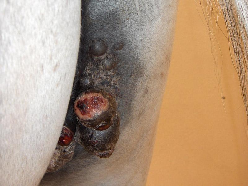
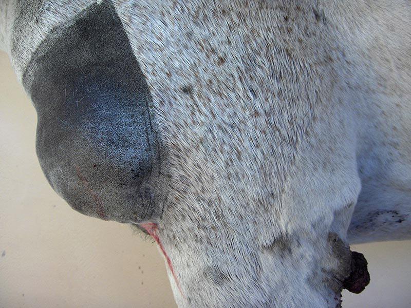
- Fibroblastic sarcoids with fleshy and ulcerated with local infiltration (figure 2)
- Mixed sarcoids present with any or all of the types above (figure 1)
- Malevolent sarcoids are aggressive, invasive tumours that proliferate rapidly and may spread along fascial planes and vessels. Malignant sarcoids being extremely rare
💡 Sarcoids can develop in any location, either as a single tumour or as multiple tumours of different types. The most common locations for sarcoid development include the head, neck, extremities and ventrum
Diagnosis
A presumptive diagnosis can often be made based on clinical appearance, but histopathology is necessary for a definitive diagnosis. An experienced pathologist is required to distinguish a sarcoid from a fibroma or fibrosarcoma and make an accurate diagnosis.
⚠️ Be aware that the trauma and irritation caused when taking a biopsy can result in the sarcoid becoming bigger (proliferation)
Treatment
Although sarcoid tumours do not metastasise, they can significantly impact the function and appearance of patient depending on the tumour location, size and numbers. Inappropriate treatment can irritate them and make the situation worse. Unfortunately, there is currently no completely effective treatment plan. Treatment options include
Surgical management
Physical excision includes conventional surgical removal and/or carbon dioxide (CO2) laser excision The chosen technique must remove the mass with a 2-3 cm margin to prevent recurrence and spread. It is often difficult to achieve such wide margins, hence CO2 laser excision is preferred as it causes delayed cell death beyond the margin achieved at surgery. If conventional incision is used, associating additional treatments should be considered.
💡’Banding’ - using elastic bands or a handheld elastrators - is not advisable as there is no guarantee that clean/wide margins can be removed
Cryotherapy
Therapy with liquid nitrogen can achieve cell membrane disruption and death by applying repeated freeze/heat and thaw/cooling cycles. This process does not have a great depth of action but is suited in combination with conventional excision to widen the margins of the surgical site.
Radiotherapy
Radiotherapy kills cells by damaging their DNA and RNA. This is an expensive process which requires specialist equipment not suitable for common field treatments.
Chemotherapy
Chemotherapy can be via injectable cisplatin, topical five-fluorouracil (5-FU) or a compounded topical cream containing 5% fluorouracil, heavy metals and thiouracil (known as AW3/4-LUDES2 cream). It can cause extensive skin lesions which can predispose the horse to other issues such as pain, inflammation and secondary infection.
⚠️ Extreme care must be taken when handling these drugs as they are cytotoxic and carcinogenic to people; correct PPE (personal protective equipment) such as special disposable chemoprotective gloves, disposable protective clothing, and eye and face protection must be worn and recommended procedures followed
Immunotherapy which stimulates the a local cell-mediated immune response inducing cytotoxic T cell and natural killer cell activity against tumour cells. A common immunomodulator used is Bacillus Calmette and Guerin (BCG), an attenuated strain of Mycobacterium. Other topical immune modulation and antiviral agents used have variable degrees of success and include herbal, compounded creams containing thuja, bloodroot and zinc chloride, as well as antiviral drugs used to treat herpesvirus-induced skin lesions in humans (Acyclovir).
Prognosis
This is always guarded as there is no gold standard treatment and each case can respond and evolve differently. Every failed attempt at treating the disease reduces the overall success rate by 50%. Therefore, careful consideration to the treatment and frequent follow up of cases is very important to cure the disease.
Prevention
Avoiding fly contamination and treating and covering wounds promptly can help reduce or even prevent the spread of the disease. Appropriate and timely intervention can also avoid the spread and progression.