

Hoof
The hoof or foot is a complex structure made up of different parts: the keratinised external capsule (forming the sole and hoof wall), the internal bones (the third phalanx or coffin bone, second phalanx or pastern bone, and the distal sesamoid or navicular bone), and numerous soft tissue structures.
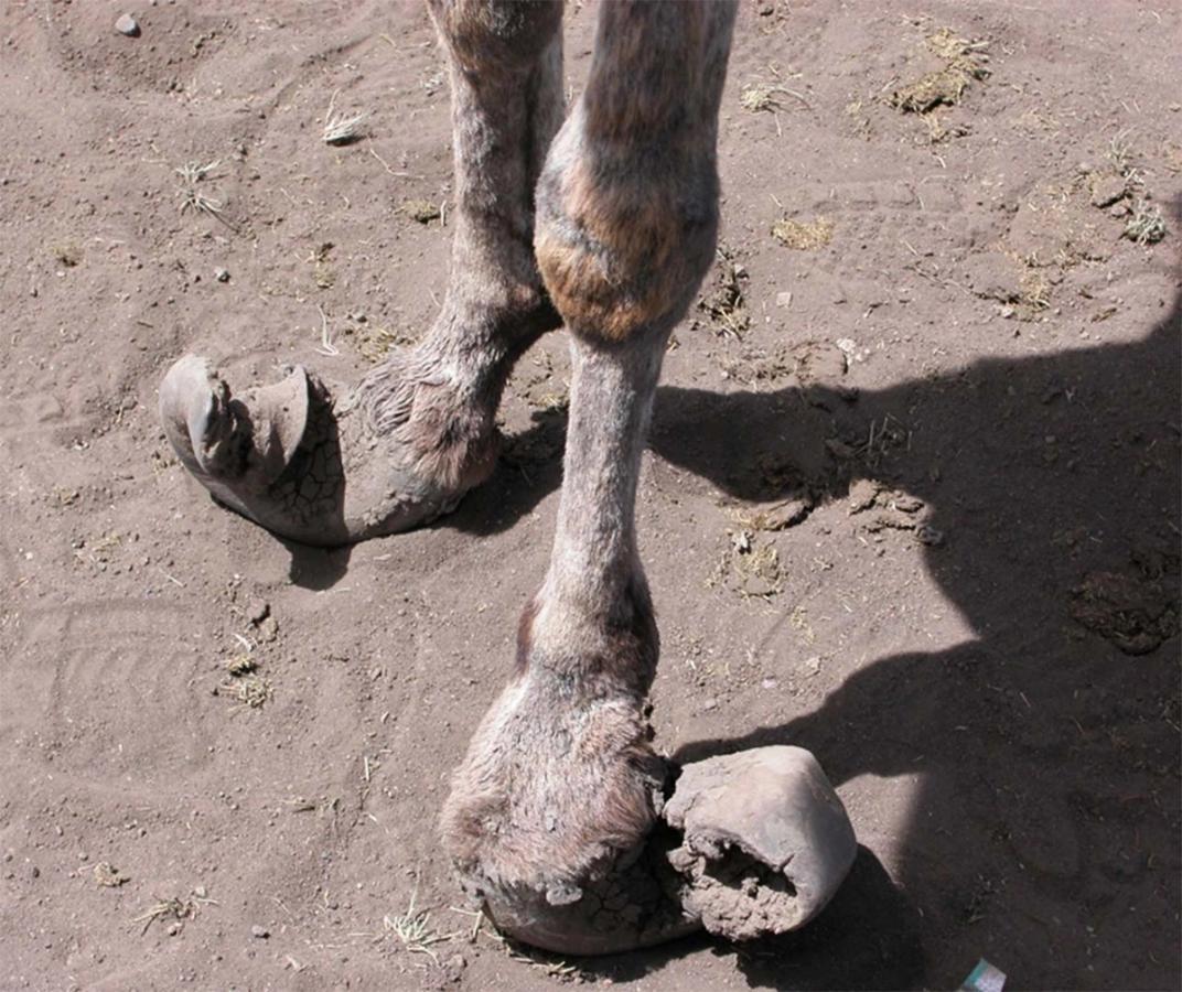
Components of a hoof
The keratinised hoof wall joins with the distal limb at the coronary band - a juncture between the skin and hair, and the hoof wall. The hoof wall then joins at the bottom of the foot with the sole and frog, at the white line and bars. The hoof wall does not have nerves or blood vessels; it's a continually growing, keratinous material that needs to be trimmed or be naturally worn off. The hoof wall is relatively inflexible and cannot expand when an injury or disease causes the structures inside to enlarge/swell. As such, injuries causing fluid accumulation, such as a haematoma or abscess will cause painful internal pressure to the hoof structures, that is manifested as severe often toe touching lameness.
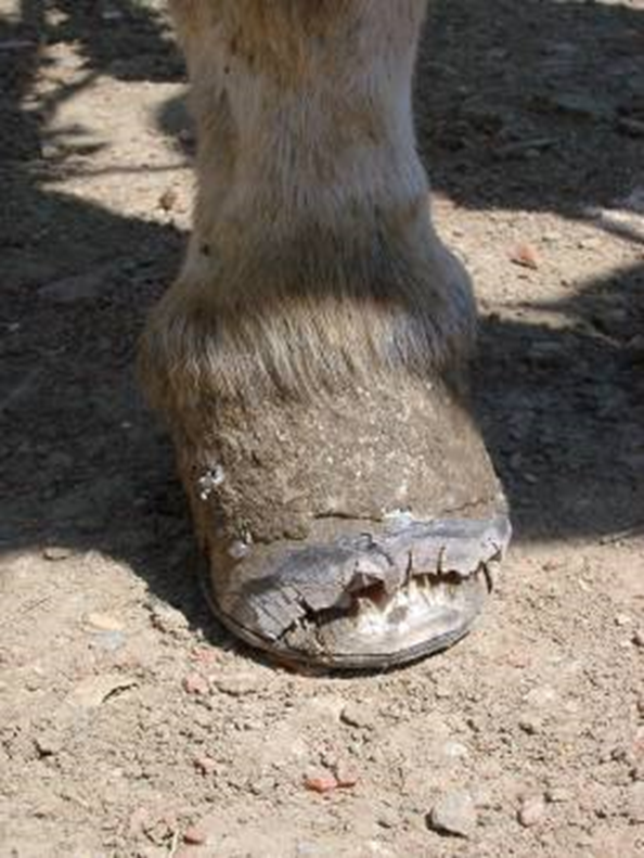
📚 The hoof wall grows a rate of approximately 0.25 inches per month. Healthy hooves should not have cracks or rings. These can make internal structures vulnerable and indicate wider systemic issues such as poor nutrition or severe inflammation of the hoof horn — a condition known as laminitis
The bones inside the hoof articulate to create the proximal interphalangeal joint, or ‘pastern’ joint (between the first and second phalanx, also referred to as P1 and P2 ), and the distal interphalangeal joint, or ‘coffin’ joint (between the second and third phalanges, also referred to as P2 and P3).
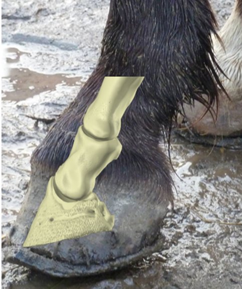
Soft tissue structures within the hoof provide both stability and controlled movement of the joints. The main soft tissue structure of clinical relevance in the hoof is the deep digital flexor tendon, which courses down the back of the limb and over the navicular bone, to attach to the back of the coffin bone (or P3). Other soft tissues structures worth of notice are:
- The collateral ligaments, always located both medially and laterally to the joint space (and unique, as they are intraarticular, but extrasynovial);
- The distal sesamoidean, or impar ligament;
- The proximal sesamoidean, or T ligament of the navicular bone — both of the latter two having recently been found to cause lameness in large numbers of sport horses (since the widespread use of advanced diagnostic tools such as MRI).
Synovial structures within the hoof include pastern joint, the coffin joint, and a separate navicular bursa. This is a fluid-filled pouch that sits between the navicular bone and the deep digital flexor tendon, and that helps cushion and protect the bone and tendon.