

An average adult horse can weigh close to 500kgs, yet the average size of a single limb of a horse does not hugely differ in diameter to that of a human's. Horse's joints are therefore designed to withstand a huge amount of weight on a very small surface. Long-term, however, this predisposes them to wear and tear of the cartilage, with secondary issues such as osteoarthritis, fractures and soft tissue damage occurring commonly.
A joint is composed of a number of components such as bones, ligaments, muscles and sesamoid structures, all working towards two major functions: (1) to enable movement, and (2) to transfer load in an energy-efficient manner. Normal joint movement is pain-free, follows a predictable range of movements, is controlled by associated soft tissue structures (namely nerves, muscles and tendons), and occurs in an almost frictionless manner.
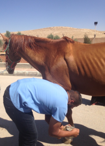
Joint disease affects most commonly high motion and high pressure joints such as the distal interphalangeal (or coffin) joint, the metacarpo-phalangeal and metatarso-phalangeal (or fetlock) joints, the carpi (or knees), the tarsi (or hocks), and the stifle joints (or true knees).
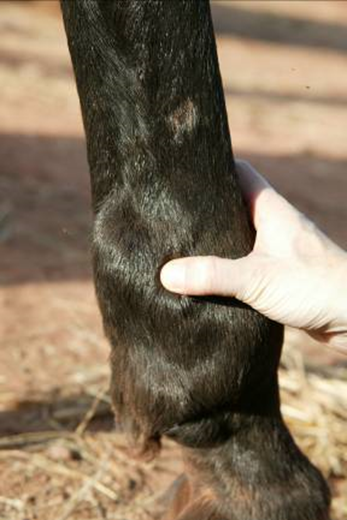
The most common signs of joint disease include loss of mobility and pain (visible as asymmetric gait or lameness), and joint distension or thickening. Clinical signs of joint disease can be exacerbated so as to be visible by the attending physician, by compressing and/or stretching the joint with the common clinical practice of "flexion tests"
Components of a joint
Joints are the point of connection between two or more bones. Most joints are mobile, although not all (for example, the tarso-metatarsal joint). Components of a joint include:
- Joint capsule. The joint capsule is a thick fibrous shell which is lined by a thin subsynovium (lamina propria) and the synovium (synovial membrane). The intima (or innermost) layer of the synovium is constituted by synoviocytes, which produce synovial fluid. The synovial fluid is colourless to pale-yellow and it is an ultrafiltrate of plasma containing hyaluronan and lubricin. Its function is to occupy and lubricate the intra-articular space.
Equine synovial fluid is rich in mononuclear cells (90% of the total cell count in joint fluid) with the remainders of the cells being polymorphonuclear leukocytes. A normal sample contains fewer than 500 nucleated cells/µL.
- Ligaments — which are tough, yet moderately elastic bands of fibrous tissue.
- Meniscus — a cartilage cushion between two bones.
- Articular cartilage, which is the principal component of any joint, allowing concurrent motion and weight bearing with minimal friction. The vast majority of articular cartilage is composed of type II collagen (85-90%), proteoglycans, and chondrocytes. Although the chondrocytes represent only a small volume of the cartilage, they maintain and turnover the entire extracellular matrix.
- Subchondral bone lies directly below the articular cartilage. While histologically similar to bone in other locations, it is thinner than cortical bone and its haversian system is orientated parallel to the joint surface, rather than parallel to the long axis of the bone (as would be for diaphyseal cortical bone). Subchondral bone is highly pliable, and its deformability exceeds that of the diaphyseal cortical shaft, as it has the important function of force attenuation.