We use cookies
We use cookies to ensure that we give you the best experience on our website. Would you like to accept all cookies for this site?
Intravenous fluid therapy (IVFT)
Intravenous fluid therapy (IVFT) is used to treat two conditions, which each have a very different approach and purpose.
Shock (except cardiogenic shock). IVFT is used in the treatment of shock where there is impaired oxygen delivery to tissues due to blood or fluid loss (hypovolaemic), physical obstruction of blood flow to the heart (obstructive) or maldistribution of blood flow (distributive). In this instance IVFT is urgent and aggressive to increase blood pressure, cardiac output and perfusion of organs and tissues.
Dehydration. Dehydration occurs through overall loss of fluid from the body, which is not replaced through drinking over a prolonged period of hours. This more gradual loss of fluid results in loss of interstitial and intracellular fluid to maintain intravascular fluid volume. The approach of IVFT in this instance is to initially treat hypovolaemic shock, followed by the replacement of fluid over a 24 hour period.
Which solution?
0.9% sodium chloride or compound sodium lactate (CSL) (also called Hartmann's solution) are acceptable for use in most situations for the treatment of shock and dehydration. Importantly do not mix CSL with blood products or sodium bicarbonate. For longer term maintenance and treatment of certain metabolic conditions, other solutions should be considered.
Treating shock
Shock is a life-threatening condition and must be treated as an emergency. Before treating with IVFT you must evaluate the cardiovascular system. This will inform your approach to IVFT as aggressive IVFT in animals with cardiac disease will increase demand on an already impaired heart and risks causing or worsening congestive heart failure.
The aim of IVFT for treatment of hypovolaemic shock is to increase the intravascular volume and therefore perfusion of tissues. This is done through the delivery of boluses of fluid over a short period of time, followed by re-evaluation of the animal for improvement in cardiovascular parameters. If signs of shock persist, repeated fluid boluses are indicated (see below).
Examination of the following physical examination parameters is important to guide IVFT treatment for shock:
Pulse rate. Assess pulse rate by palpating the femoral pulse. Tachycardia is a common sign of shock. See the Quick Reference for basic physiology for normal heart rates.
Pulse quality. As well as evaluating pulse rate from the femoral pulse, you can also evaluate pulse quality. In the early stages of shock, pulses will be 'bounding' (hyperdynamic) - these feel like a stronger, shorter pulse than normal as cardiac output increases to try to maintain blood pressure. As hypovolaemic shock progresses pulses become weaker, but remain short (hypodynamic). Palpation of the metatarsal pulse can enable more sensitive detection of abnormalities than the femoral pulse, however is a skill which needs practice to get used to what is normal (Figure 1).
Mucous membrane (MM) colour. Mucous membrane colour provides information about perfusion. In hypovolaemic shock MM will be pale, whilst in distributive shock (e.g. septicaemia) they can be bright red (congested).
Capillary refill time (CRT). Capillary refill time should be less than 2 seconds, however prolonged CRT is a sign of hypovolaemic shock (> 2 seconds) and can be very brief in distributive shock (< 1 second) where the mucous membranes are congested.


Figure 1 - Evaluation of pulse quality and rate can be done through palpation of the femoral (left) and metatarsal pulses (right). The metatarsal pulse is a sensitive indicator of changes in an animal's haemodynamic status, however takes practice to get familiar with what is normal.
Calculating shock IVFT rate
Here we will describe the use of isotonic crystalloids for the treatment of hypovolaemic shock, as is the case for intra-operative haemorrhage or haemorrhage following trauma.
Hypertonic crystalloids or colloid solutions fluids can be used for the treatment of shock and have benefits in requiring the administration of smaller volumes to achieve the same expansion of intra-vascular volume, however they need to be used with careful evaluation of blood electrolytes and patient hydration. Most patients can be treated safely and effectively in resource limited settings using isotonic crystalloid solutions, such as 0.9% NaCl (normal saline) or Compound Sodium Lactate (CSL).
Shock bolus. Give 20ml/kg fluid bolus over 15 - 20 minutes and then evaluate shock parameters for improvement. If signs of shock persist, give another 20ml/kg bolus over 15 - 20 minutes and re-evaluate signs. This process can be repeated until an improvement is seen, up to a total dose of 90ml/kg. It is important to address the underlying cause of shock as quickly as possible, for example preventing ongoing blood loss either externally as in the case of a haemorrhaging wound or internally as in the case of a ruptured splenic mass.
Table for crystalloid bolus infusions to be administered over 20 minutes before re-evaluating shock parameters
| Weight (kg) | Crystalloid bolus over 20 minutes |
|---|---|
| 5 | 100ml |
| 10 | 200ml |
| 15 | 300ml |
| 20 | 400ml |
| 25 | 500ml |
| 30 | 600ml |
When delivering fluid boluses you will likely need to place the largest gauge IV catheter possible to achieve adequate infusion rates. This can be a challenge in collapsed patients and minimising failed attempts is key for preserving IV access. In large patients IV fluid infusion through catheters in both cephalic veins may be needed.
Treating dehydration
Estimating dehydration
Estimation of how severely dehydrated a patient is helps us to calculate replacement of this through intravenous fluid therapy over a 24 hour period.
Physical examination components used to assess hydration level are:
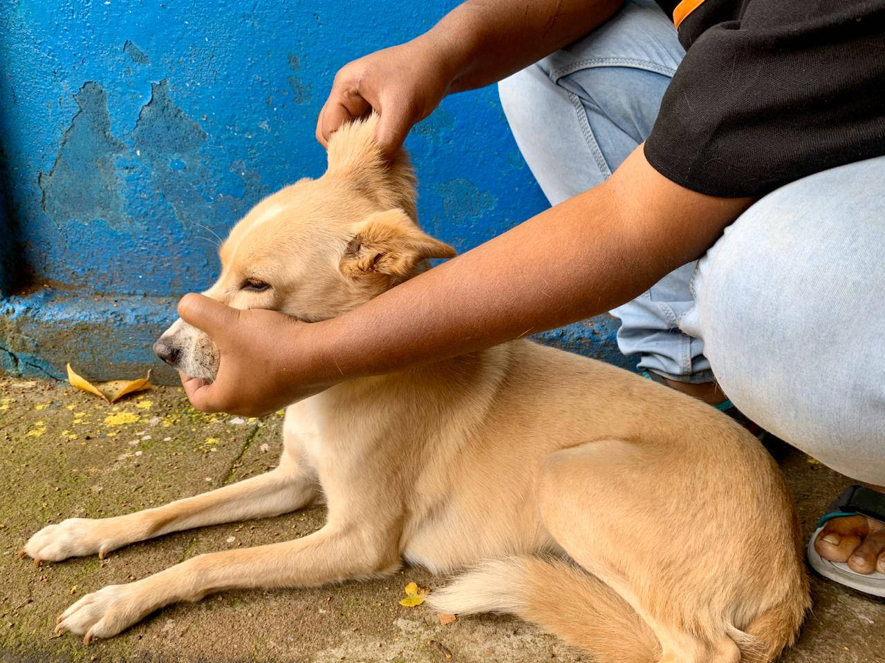
Skin elasticity. This can be evaluated by gently pinching the skin to the back of the head. Loss of skin elasticity due to dehydration results in the skin remaining tented for a time after pinching.
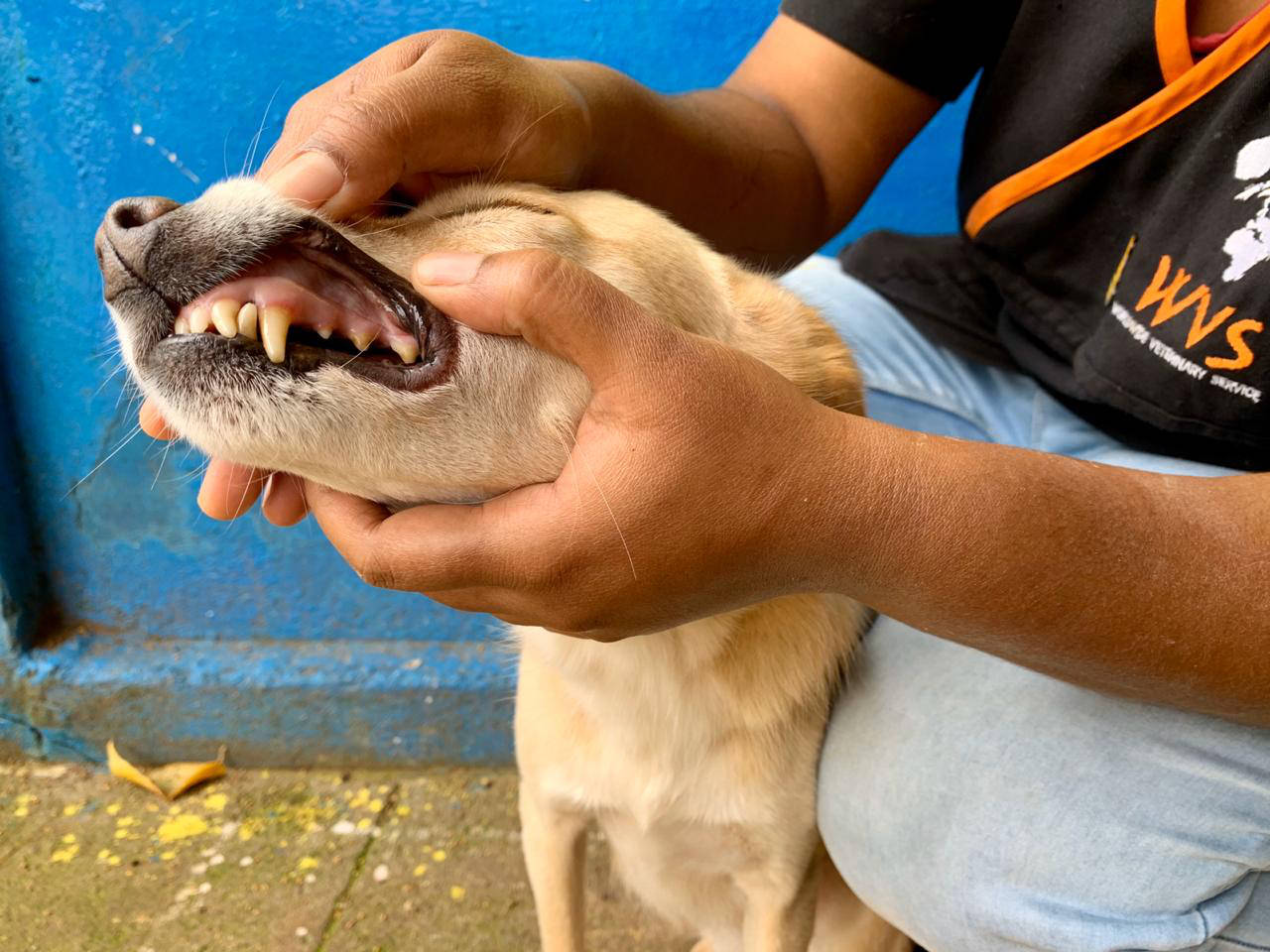
Oral mucous membrane (MM) moisture. Evaluated by observing or touching the mucous membranes. They should be moist to touch. Dehydration results in tachy or dry mucous membranes.
Capillary refil time (CRT). Evaluated by gently pressing the gum and releasing whilst observing return of the pink colour. In a normal animal, the blanched area will return pink in under 2 seconds, however this becomes prolonged with dehydration.
Pulse rate and quality. As dehydration progresses signs of hypovolaemic shock become apparent, including tachycardia, weak pulses and cold extremities (see above).
Eye position. As animals become more severely dehydrated, the eyes become sunken in the sockets due to dehydration of the periorbital fat pads.
Mentation. Assess the animals awareness of its surroundings and state of consciousness. In severe dehydration the animal will become subdued and eventually unable to stand.


Figure 2 - Assessment of the moisture and colour of oral mucous membranes (left) and skin elasticity (right) are used in the physical examination to provide information about hydration status.
Physical examination and percentage dehydration
| Clinical assessment | Estimated percentage dehydration |
|---|---|
| No clinical signs | < 5% |
| Mild skin tent | 5 - 6% |
| Definite skin tent, dry gums, mild increase in CRT, slightly sunken eyes. | 6 - 8% |
| Significant skin tent, dry oral MM, increased CRT, sunken eyes, signs of hypovolaemia (see above) | 8 - 10% |
| Severe skin tent, dry oral MM, obvious sunken eyes, obvious signs of hypovolaemia, subdued mentation,possible recumbency | 10 - 12% |
| Hypovolaemic shock, moribund, death imminent | 12 - 15% |
Calculating replacement IVFT requirements
An animal's 24 hour fluid provision can be calculated as:
Maintenance requirement + Replacement for dehydration
This is divided by 24 to give an hourly IV fluid rate.
Maintenance requirement: 50-60ml/kg/day
Dehydration replacement (ml): Body weight x percentage dehydration x 10
The tables below give examples for calculating the infusion rate for various body weights. When setting infusion rates based on drips per second it is important to check 'drips per ml' for the infusion set you are using. Here we use a standard set of 20 drips per ml.
Table for IVFT rate for dogs estimated to be 5% dehydrated
| Body weight | Hourly amount (ml/hour) | Maintenance (ml) | Replacement (ml) | Total 24 hour requirement (ml) | Infusion rate |
|---|---|---|---|---|---|
| 5kg | 23 | 300 | 250 | 550 | One drip every 8 seconds |
| 10kg | 46 | 600 | 500 | 1100 | One drip every 4 seconds |
| 15kg | 69 | 900 | 750 | 1650 | One drip every 3 seconds |
| 20kg | 92 | 1200 | 1000 | 2200 | One drip every 2 seconds |
| 25kg | 115 | 1500 | 1250 | 2750 | One drip every 2 seconds |
| 30kg | 138 | 1800 | 1500 | 3300 | One drip every second |
| 40kg | 183 | 2400 | 2000 | 4400 | One drip every second |
| 50kg | 229 | 3000 | 2500 | 5500 | One drip every second |
Table for IVFT rate for dogs estimated to be 10% dehydrated
| Body weight | Hourly amount (ml/hour) | Maintenance (ml) | Replacement (ml) | Total 24 hour requirement (ml) | Infusion rate |
|---|---|---|---|---|---|
| 5kg | 33 | 300 | 500 | 800 | One drip every 5 seconds |
| 10kg | 67 | 600 | 1000 | 1600 | One drip every 3 seconds |
| 15kg | 100 | 900 | 1500 | 2400 | One drip every 2 seconds |
| 20kg | 133 | 1200 | 2000 | 3200 | One drip every 1.5 seconds |
| 25kg | 167 | 1500 | 2500 | 4000 | One drip every second |
| 30kg | 200 | 1800 | 3000 | 4800 | One drip every second |
| 40kg | 267 | 2400 | 4000 | 6400 | 1.5 drips every second |
| 50kg | 333 | 3000 | 5000 | 8000 | 2 drips every second |
© WVS Academy 2025 - All rights reserved.