

Timing of collection and analysis
Collecting a sample early in the morning, particularly the first urination of the day, usually provides a more concentrated sample, which is particularly useful for evaluating the kidneys’ concentrating ability. It also yields a higher concentration of cells, bacteria and casts.
If possible, collecting more than one sample at different time points can also be useful, particularly for parameters such as specific gravity, which is likely to vary, depending on hydration status. Glucosuria may be more obvious in a post-prandial sample (i.e. after feeding).
Urinalysis should be performed as soon as possible following collection, within an hour ideally. Over time, changes to pH, glucose, ketone and bilirubin levels can occur, cells and casts degenerate and crystals may dissolve or precipitate. If samples have to be stored, they should be refrigerated, then warmed back to room temperature and mixed prior to being tested. Bacteriology should ideally be carried out with 24 hours of sample collection.
Free-catch collection
Advantages: This method is easy to perform, cost-effective, and it can also be carried out by owners at home.
Disadvantages: Samples are contaminated with bacteria from the genital tract and environment, so cannot be used for culture and sensitivity testing.
Method: For dogs, hold a kidney dish or collection pot underneath during normal urination. For cats, non-absorbent litter may be used, with a syringe or pipette used to remove the urine. Manual expression should generally be avoided due to risk of rupture and iatrogenic damage.
Tips: It’s a good idea to remind owners to avoid re-using containers which have previously contained sweet foods such as jam, honey etc., even if they have been cleaned. Trace amounts of glucose can remain and cause a false positive on analysis. The middle portion of the stream of urine should be collected, as this is less likely to be contaminated with material from the genital tract.
Cystocentesis
Advantages: This method results in a sterile sample by avoiding genital tract contamination, meaning it can be used to test for culture and sensitivity.
Disadvantages: This may introduce blood into the urine sample due to entering the bladder via needle (iatrogenic haematuria). If the patient moves during the procedure, this may result in trauma; and if the needle pierces the large intestine by mistake, this can result in bacterial contamination. Cystocentesis must be carried out by a veterinarian, and is more technically difficult than free-catch.
Method: This procedure should be performed using aseptic technique. Patients may be in dorsal/lateral recumbency, or standing.
Equipment:
- Needle selection depending on size of animal: 5/8 inch or 1/2 inch needle (21-23G in dogs, 23G in cats)
- Syringe (e.g. 5-10ml)
- Plain collection tube (sterile if using for culture)
The urinary bladder is located in the caudal abdomen, just cranial to the pelvic brim and ventral to the colon. Identify by gentle palpation and then stabilise it using one hand. If it cannot be palpated, it is likely that there is not enough urine volume to perform the procedure at this time.
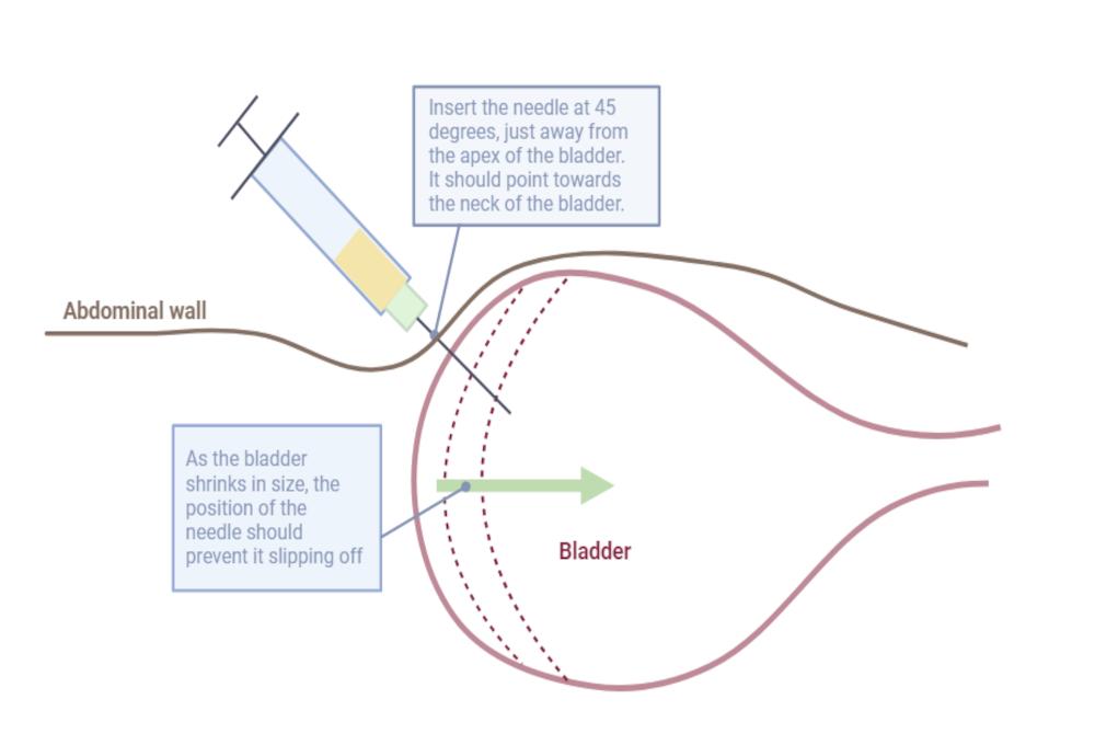
In a recumbent animal, insert the needle on the midline, just cranial to the pelvic brim. In the standing animal a right sided approach reduces the risk of penetrating the descending colon. The needle should be at a 45 degree angle to the body wall, aimed in a caudal direction, towards the neck of the bladder, and should enter a few centimetres away from the apex. This means that as the bladder deflates, it is less likely to slip off the end of the needle.
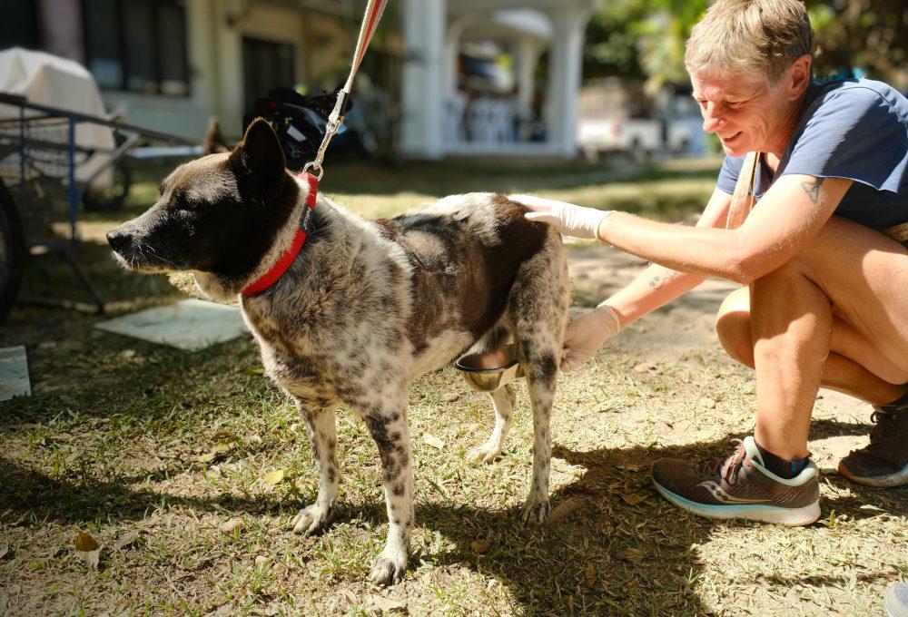
Figure 3. Performing cystocentesis on a female canine patient.
Tips: Using an ultrasound will allow you to visualise the bladder and needle as you take the sample.
The hyperechoic (white) spot at the top of the screen is the tip of the needle as it is inserted into the bladder. Watching the screen for the correct placement of the needle before drawing back on the syringe can help ensure safe sample collection.
In some circumstances, such as with large or nervous dogs, the standing approach may result in less stress for the patient and could provide an alternative option to the recumbent positions (1). The urinary bladder is entered through the lateral abdominal wall; in bitches, this is around the level of the caudal mammary glands and, in males, just lateral to the midline after reflecting the prepuce away from you.
Cystocentesis is contraindicated where there is a suspected mass in the bladder, due to risk of seeding tumour cells in other areas of the abdomen, as the needle is withdrawn. It should also be avoided in cases of severe bladder disease (due to increased risk of rupture), in female dogs with suspected pyometra due to the risk of piercing the infected uterus, and in animals with clotting disorders. Where body fat impedes palpation and stabilisation of the bladder, it may not be safe to perform the procedure.
Catheterisation
This method involves advancing a catheter through a patient’s urethra to reach the bladder. It is rarely indicated as a primary method of urine collection, due to the risk of bacterial contamination from the genitalia causing an infection higher up the urinary tract, as well as potential pain associated with the procedure. However, samples may be collected in this manner if other methods are not possible, or if a catheter is already in place for other reasons.