

A number of large blood vessels (called direct cutaneous arteries and veins) supply the skin. Each region of skin supplied by a single artery is called an angiosome. The locations of many direct cutaneous arteries and their angiosomes have been mapped out in dogs, but there may be individual variations. There is less available information on known angiosomes in cats. There are known differences between the species, so anatomy of the dog is not automatically transferrable to the cat. (This will be discussed further in later modules).
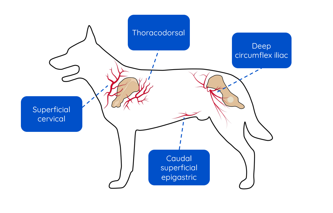
Direct cutaneous arteries emerge from the skeletal muscle layer, before entering the subcutis, including through the cutaneous trunci muscle (in areas of the body where it is present).
In the dermis layer of the skin, the vessels branch further into a capillary network, creating an area of a dense vasculature. The dermis therefore has the richest blood supply in the skin (compared to the subcutis and epidermis). Capillary loops in the dermis pass close to the epidermis layer but do not enter. The epidermis receives nutrients and oxygen via diffusion, instead.
The branching vessels in the subcutis and dermis layers of the skin form three connected networks of vessels called:
- Superficial plexus
- Middle plexus
- Deep/ subdermal plexus

The dense branching vessels in these plexuses form extensive connections within the skin. This means there is good collateral blood flow, i.e. alternate routes for blood flow outside of the main vein or artery.
Blood supply and wound healing
A good blood supply is crucial to healing. Evaluation of blood supply is an important factor in clinical decision making and will be revisited in later modules. A compromised blood supply can lead to chronic or non-healing wounds, and tissue may even become necrotic (dead).
If a wound requires surgical closure, but primary closure would result in unacceptable tension across the wound, techniques such as undermining of the skin, or skin flaps, may be required. When using these techniques, it is important to ensure any skin which is operated on maintains an adequate blood supply. When undermining the skin, you should dissect below the subdermal plexus and try to preserve direct cutaneous vessels to avoid disrupting cutaneous blood supply. This will help to reduce the risk of wound dehiscence and skin necrosis. In the distal limb, the plane of dissection should be between the fat and the deep fascia covering the skeletal muscle. Dissection below cutaneous muscle is required in areas where this is present i.e. head, neck, thorax and abdomen.
Subdermal plexus flaps are flaps of skin used in reconstructive surgery whose blood supply relies solely on the residual subdermal plexus connections between the base of the flap and the adjacent skin. Axial pattern flaps are skin flaps that incorporate the larger direct cutaneous vessels, and so these skin flaps have a more robust blood supply. It is estimated that an axial pattern flap can survive being 50% larger than a subdermal plexus flap due to this more robust blood supply.
Your choice of surgical flap will be governed by many factors, including available blood supply: this will be covered in more detail in later modules.
Innervation
The skin has a rich nerve supply which responds to touch, pressure, temperature, nociception (pain sensation) and pruritis (itch). The skin is also innervated by the autonomic nervous system with motor neurones that control vasoconstriction and vasodilation, as well as the activity of sweat and sebaceous glands.
Analgesia (pain relief) is an important component of wound management and will be covered throughout this course.