

Patient preparation
Withhold food. For at least 6 hours prior to radiographs. This ensures the stomach and intestines have had time to empty, which allows for increased contrast of abdominal organs. Water may be given to prevent dehydration.
Allow defecation and urination. Again, encouraging an empty digestive tract will increase the contrast of all abdominal organs, meaning a better radiograph.
Clean coat. Dirt should be cleaned off and the coat should be dry.
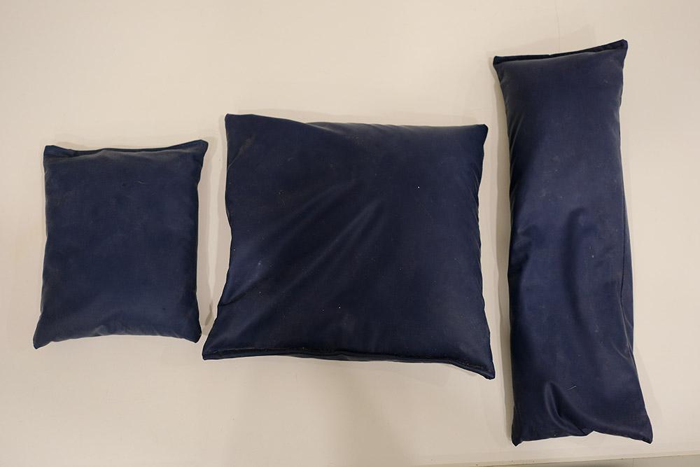
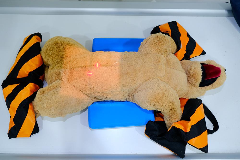
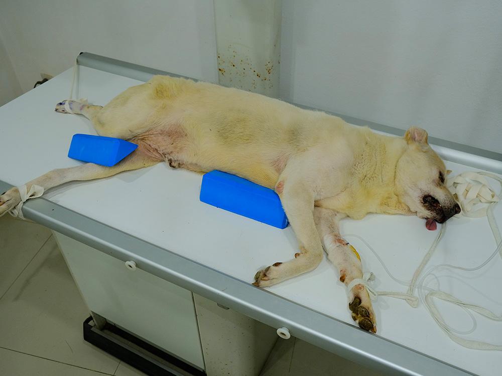
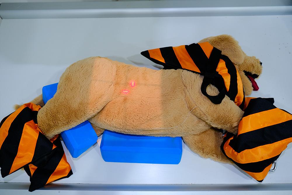
Positioning aids. These should be used to keep patients in place, rather than staff holding the patient.
Sedation/ general anaesthetic. Sedation is necessary where the patient is unlikely to remain still. It also allows us to use positioning aids more easily rather than manual restraint. Ensure a thorough clinical exam is carried out before giving sedation.
Standard radiographic views
For abdominal radiographs, the standard views are:
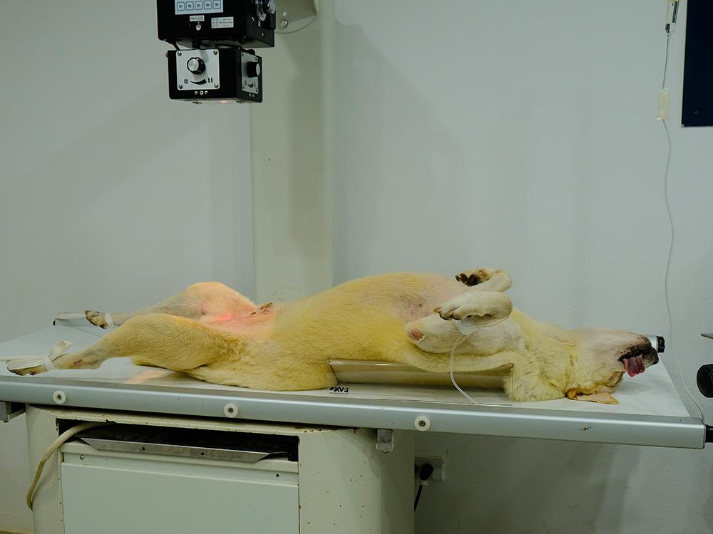
Ventrodorsal. The patient is in dorsal recumbency, on their back with their legs in the air. Collimation cranially is to the level of the last rib/ diaphragm, caudally to the greater trochanter of the femur, and laterally to the skin edges.
Right and left lateral. The patient is on their side, with the corresponding side to the plate. Collimation cranially is again to the level of the last rib, cranial to the liver. Collimation caudally is again to the level of the greater trochanter.
In certain circumstances, the clinician may opt for a dorsoventral view as well, to maximise information. They may also only take one instead of two lateral images, for dyspnoeic patients, to minimise time spent in lateral (which compromises breathing). We may also do one lateral in patients that are becoming more stressed out as the procedure goes on.
Exposure considerations
Low kV, high mAs. The abdomen is less mobile than the thorax, so there is less concern for motion blur and a longer exposure time may be used. As the abdominal organs are similar in consistency, high penetration is not required, so we can use a low kV. However, we must increase contrast as similar consistency organs are difficult to distinguish, so a high mAs is opted for.
End expiratory view. The radiograph is taken at the end of expiration, as there is less movement at this point, and so there's a lower chance of movement blur occurring.
Use a grid. In patients with abdomens deeper than 10cm, a grid is placed on the plate to reduce scatter radiation.
Safety considerations
Before performing radiography, ensure the warning light is turned on so that all staff are aware that harmful X-ray radiation is being used and they may then choose to vacate the area. Sedation and positioning aids should be used, so all staff may stand behind the protective lead shielding screen during X-ray exposure. In certain specific emergencies, staff may use manual restraint, so long as appropriate PPE is donned, including lead gloves, lead aprons, and thyroid shields. All staff involved should also wear dosimeters to allow monitoring of radiation doses.