

The clinician must learn when abdominal radiographs are appropriate, or whether a different diagnostic technique may yield more conclusive results.
Common indications for abdominal radiographs include:
Abdominal distension. This may suggest free fluid, masses, pyometra, or a number of other conditions which may be diagnosed with radiographs.
Investigation of masses. If we have palpated a mass, radiographs allow us to survey the body for metastases such as within the thorax or lymphatic system. This, alongside a fine needle aspiration (FNA), will give us an insight into the prognosis and possible treatment options. If markedly elevated lymphocytes are present on haematology, and lymphoma is suspected, screening radiographs may also be taken which include the abdomen.

Weight loss. This is non-specific and may result from a variety of conditions. With radiography, we may diagnose causes such as neoplasia, pancreatitis, and renal or hepatic causes of weight loss. Ultrasonography is more sensitive to diagnosing conditions such as inflammatory bowel disease (IBD), or organ dysfunction (ileus).

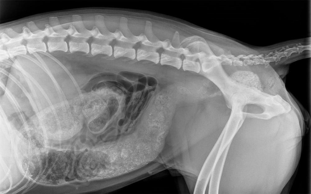
Abdominal pain. Again, an unspecific finding, however, radiography may give an insight into any physical causes of pain. For example, distended loops of intestines, foreign body or faecal impaction.
Trauma. Following stabilisation of trauma patients (e.g. road traffic accident), we may radiograph the abdomen to check for internal injuries (diaphragmatic herniation, ruptured bladder, or bleeding). This will guide the treatment plan.

Gastrointestinal signs. For repeatedly vomiting patients, we may suspect a foreign body or gastrointestinal blockage. Radiographs may be used in conjunction with a detailed history and potentially an ultrasound exam to confirm this.

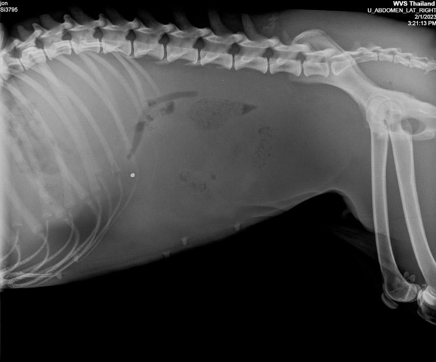
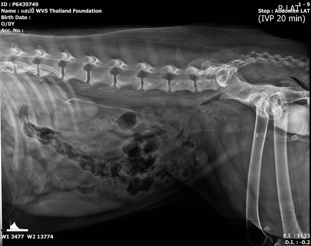
Urinary signs. We may use radiographs to assess the urinary tract for the presence of uroliths (radiopaque), tumours (transitional cell carcinoma), or structural changes that may explain urinary incontinence. Contrast radiography may be used to highlight structures and improve radiography’s diagnostic abilities.
Reproductive system assessment. Radiography of a suspected pyometra may show us a distended uterus, filled with liquid (pus). Ultrasound is the preferred diagnostic modality for pyometra, but radiography allows us to rule out other conditions at the same time, so we may perform both for a complete picture.