

Diagnostic or not?
Once we have taken the radiograph, fill in the patient details. Then review the radiograph to determine whether it is diagnostic. Assess the exposure and contrast, ensuring you can distinguish different organs from one another and there is no over- or under-exposure. Ensure the image shows the site of interest in its entirety (but collimated to minimise scatter), and that the left/ right marker is correctly positioned. Then move on to assess abnormalities.
Roentgen signs
As previously discussed, we can base our interpretations upon these signs when writing the radiographic report. This systematic approach ensures abnormalities aren’t missed, allowing us to make a diagnosis.
Size. Abdominal organs may change size due to underlying disease processes. Breed variation makes it difficult to determine where changes have occurred, however, we can compare organs with bony landmarks to assess this. The canine kidney, for example, is roughly 3x the length of the second lumbar vertebra. Increased size may be due to hypertrophy, hyperplasia, organ congestion or gastrointestinal obstruction, whereas a decreased size is common with chronic inflammatory processes, hypoplasia, atrophy and hypovolaemia.
Shape. This may change due to physiology or pathology. Neoplasms alter organs’ shape and are often recognised by their roughly spherical appearance. Inflammatory lesions (granulomas or abscesses), cysts and obstructions also cause shape changes. Foreign bodies, gastrointestinal obstructions, or fluid or gas accumulation may also cause shape changes.
Number. The absence of an organ could be due to displacement from neighbouring organs’ pathology or congenital hypoplasia of the organ itself. Congenital aplasia means the organ failed to develop in the first place but it is very rare. Extra organs may be due to accidental double exposures, or due to the enlargement of structures that aren’t normally seen (uterus/ lymph nodes). Take care not to interpret pathological structures (cysts, tumours, enlarged adrenal glands) for additional organs, like an extra kidney. Furthermore, poor positioning may obscure certain areas of the abdominal cavity, meaning we cannot identify all organs.
Opacity. Gas-filled structures such as the lungs may become more radiopaque with masses or calcification. Soft tissue may appear darker (radiolucent) with gas-producing infections, or lighter (radiopaque) with calcification such as with urinary calculi (stones). An increase in bone radiolucency may suggest cysts or lytic lesions. Metal implants show up as white due to their strong radiopacity.
Margination. Decreased organ margin clarity may indicate free fluid in the abdominal or retroperitoneal spaces, or a lack of abdominal fat (reduced contrast) due to chronic weight loss. Inflammatory lesions of articular or joint margins (osteoarthritis) also decrease margination as they disrupt the cortical margins (bone edges).
Position. An abdominal mass cause displacement of other organs, so we may notice this without visualising the mass itself. The mobile small intestine is readily displaced and so may appear in a number of positions (this is normal). Abnormal organ position may also occur following trauma. For example, cats involved in road traffic accidents often present with diaphragmatic herniation, which involves abdominal organs being forced into the thorax, tearing or rupturing the diaphragm in the process.
Organ-specific interpretation
We interpret abdominal radiographs systematically. This may be done by assessing the individual organs or organ systems from cranial to caudal. The aim is to define the lesion, including its location and appearance, and then draw conclusions on likely differential diagnoses. Some lesions are diagnostic (GDV), whereas some highlight where the problem is likely to be (organomegaly).
Liver. The liver appears in the cranial ventral abdomen, behind the diaphragm and within the costal arch. It has a curved triangular shape and is bordered by the diaphragm cranially, the stomach dorsocaudally, and the duodenum ventrocaudally. A larger size than usual (hepatomegaly), with rounded caudoventral margins and displacement of the stomach, indicate a swollen liver. The liver is proportionally larger in young dogs and cats compared to adults.
Stomach. Present in the cranial abdomen, just caudal to the liver. Its size and shape varies based on its contents. It is large following a meal, and may become distended by gas in the case of a foreign body blockage, or GDV (gastric dilatation and volvulus). GDV can be recognised by its “double bubble” appearance, which shows gas dilation on either side of a twist in the stomach. In cases where we wish to better visualise the individual stomach wall layers, we may also opt for an ultrasound scan.
Spleen. The spleen is located on the left side of the cranial abdomen, adherent to the stomach’s greater curvature. It is mobile but is best visualised on the right lateral view, as a small triangular or ovular structure ventral to the stomach or liver. We may detect splenic masses with radiography, with large ones often seen in the mid-abdomen on lateral/ventrodorsal views, with possible small intestine displacement dorsally and caudally.
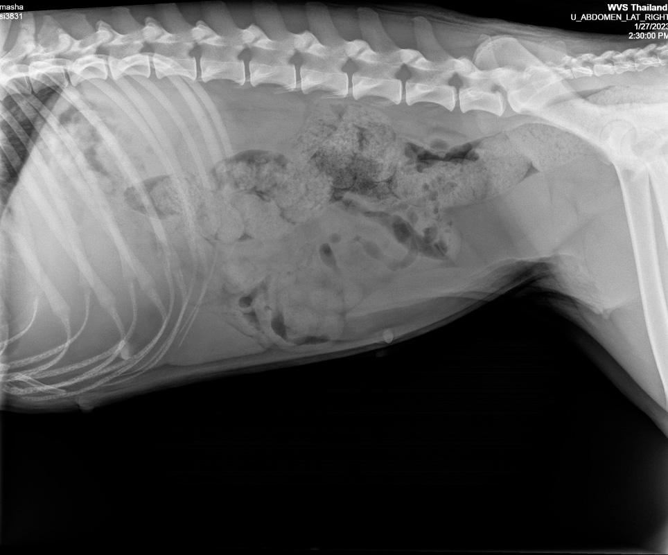
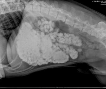
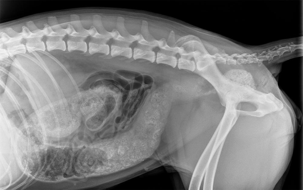
Small intestine. Radiography is not the best modality to image the small intestine. Ultrasound is more specific and lets us assess individual layers; helpful for diagnosing intussusceptions. However, we can gain “bigger picture” information on the small intestines with radiography. The small intestine’s loops are seen in the mid, ventral abdomen. Increased diameter by more than 1.6x the height of the fifth lumbar (L5) vertebra suggests ileus or “gut stasis” has occurred, due to a mechanical blockage (foreign body), intussusception or neoplastic process. Solid foreign bodies are radiopaque, and show up easily on radiographs. Gas dilation may occur secondary to blockage with a foreign body. However, dilation may also occur secondary to a functional problem, such as with peritonitis, enteritis or hypokalaemia.
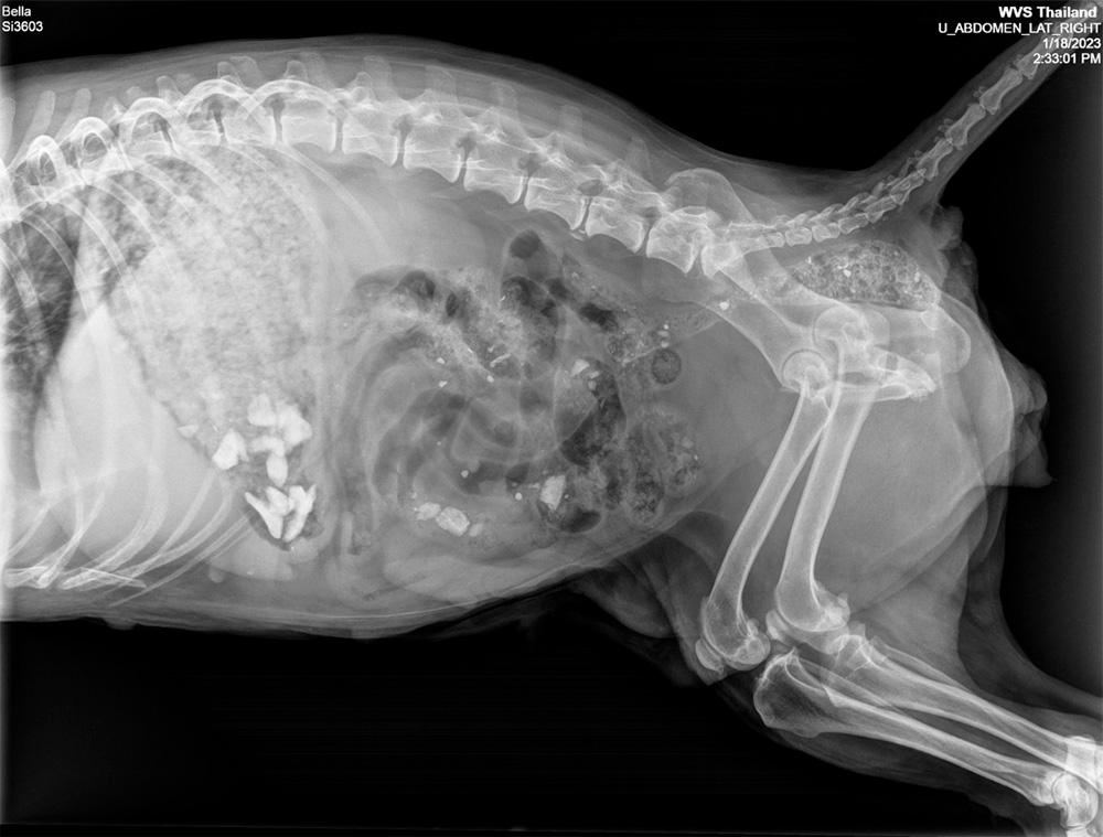
Large intestine. The large intestine is seen in the central abdomen before it passes above the pelvis to the rectum. It tends to be filled with varying amounts of faecal material, with uniform opacity. During constipation, the large intestine will dilate with radiopaque faecal material and a worrying sequela to this is megacolon. Displacement may occur ventrally due to enlargement of a kidney, local lymph nodes, or any form of neoplasia dorsal to the colon, whereas dorsal displacement follows prostate enlargement, pyometra, or an overly full bladder.
Urinary tract. This includes the kidneys, ureters, bladder and urethra. The kidneys are seen in the mid-dorsal abdomen just caudal to the stomach. The right kidney is slightly cranial to the left one. The left lateral and ventrodorsal views are best to individually visualise the kidneys. Kidneys may be abnormal in size, shape or opacity. The ureters and urethras are not visible on plain radiographs of a healthy patient, but they can be visualised using contrast radiography. The bladder is round and seen in the caudal ventral abdomen, below the colon. Uroliths (urinary stones) may be visible as areas of markedly increased opacity anywhere along the urinary tract. This is because they are crystals, and as such are solid. Transitional cell carcinoma is a neoplasm which is the most common form of soft tissue opacity visible within the bladder, often forming in the bladder’s neck.
Prostate (dog). This is located just caudal to the neck of the bladder and surrounds the urethra close to where it exits the bladder. In castrated or young dogs, this may be hard to see. In mature dogs, it can be seen as an ovular structure just cranial to the pelvic brim. Enlargement commonly causes displacement and sometimes compression of the colon or bladder, which may lead to faecal build-up within the colon. Neoplasia of the prostate occurs more commonly in castrated dogs and may result in a mineralised appearance. Benign prostatic hyperplasia is another common differential, causing enlargement of the gland. Increased size for whatever reason commonly causes.