

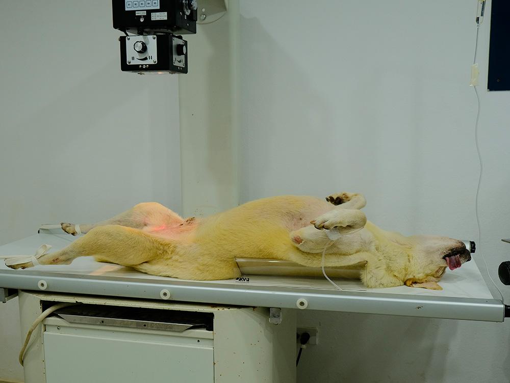
1. Position sedated patient in dorsal recumbency (spine to the plate, with legs in the air).
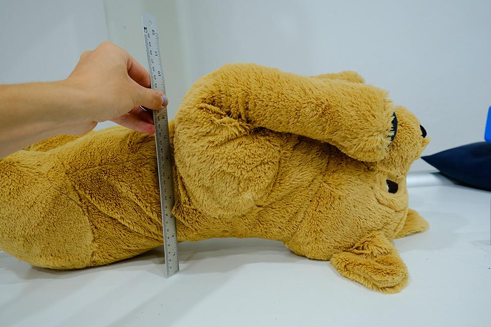
2. Measure the patient depth at the deepest point of the exposed area. If it is over 10cm, place a grid over the plate. In some X-ray tables this is not necessary (the plate is within the table in these machines).
3. Select exposure settings on the X-ray machine according to the view (VD, abdomen), and patient size.
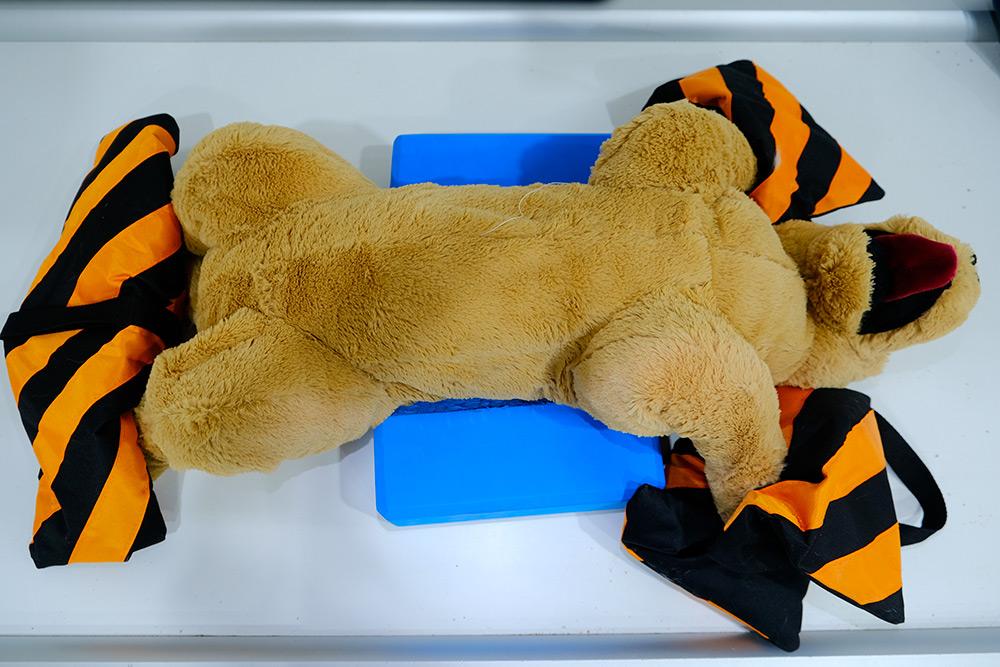
4. Place foam wedges on either side of the chest to keep the patient straight and minimise rotation.
5. Place sandbags on front and hind limbs to hold patient in position
6. Place the right/left marker within the collimation of the beam on the corresponding side of the patient. This may be added digitally.
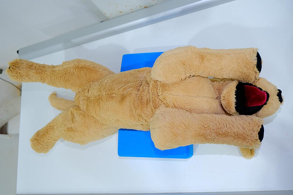
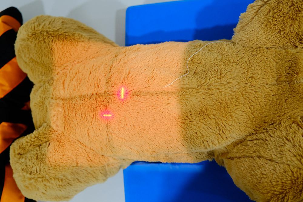
7. Centre the primary beam over the umbilicus.
8. Collimate the primary beam cranially to the diaphragm, caudally to the head of femurs, and laterally to include both skin edges.
9. Take the radiograph.