

One of the major considerations is the preparation of the patient's skin where the surgical incision will be created, referred to as the surgical site. Ordinarily, the skin is an effective barrier against invasion by many microorganisms; once the incision has been made, this barrier is breached. Both the skin and hair are a common source of infections from endogenous microbial flora, in particular, Staphylococcus spp. and Streptococcus spp. Resident microorganisms live in the cornified layers of the skin and the hair follicles, and prevention of exposure to these during surgery is critically important. An effective, pre-operative, procedure for preparation of the skin reduces the number of bacteria and therefore the risk of infection. The first step in this procedure involves clipping or shaving the surgical site. This should be done immediately prior to surgery as a longer period between clipping and surgery correlates with an increased risk of infection.
Care must be taken to avoid damaging the surface of the skin during hair removal. Damage will reduce the barrier function and encourage colonisation by bacteria.
Methods of hair removal
Regardless of the method used, always begin by shaving the incision site and moving outwards - this area is of prime importance to maximise cleanliness and keep free from skin damage.
Clipping
Clipping is the recommended method for hair removal. Electric clippers fitted with size 40 blades are suitable for this purpose. When properly maintained, clippers minimise skin trauma whilst removing hair to a very fine grade. Initially, clip in the direction of the hair growth pattern and then a second clip in the opposite direction, to ensure a smooth finish with a final cut close to the skin (Figure 1). Avoid the clippers becoming too hot as burns will cause inflammation.
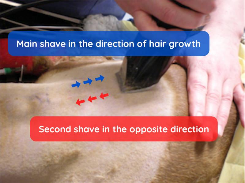
Photo credit - Mrs Denise Prisk, Dip AVN (Surgical), VTS (Anaesthesia and Analgesia), LTCL, LCGI, RVN
Clipper maintenance. Clippers should be cleaned after use by removing the blade and using a small brush e.g. toothbrush, to clean between the teeth. The addition of a small amount of oil based lubricant before use will ensure that the mechanism continues to function correctly. Clipper blades should be sharpened or replaced periodically as blunt clippers are more likely to damage the skin.
Shaving
In resource-limited settings, shaving may be the only option available; whilst previous studies have indicated that the rate of surgical site infections may increase when shaving versus clipping, there remains a lack of good-quality evidence to date. Shaving with a blade requires a much greater level of skill than it does with clippers and there is a greater risk of serious laceration if the animal moves of if the operator is not careful. The animal should be fully anaesthetised before shaving begins and operators should be supervised until competency has been demonstrated.
Sharp blade. Shaving blades blunt quickly and you are more likely to damage the skin with a blunt blade. Change blades regularly to avoid this and be sure to dispose of old blades in a sharps container.
Wet the fur. The fur is first made wet by spraying savlon solution.
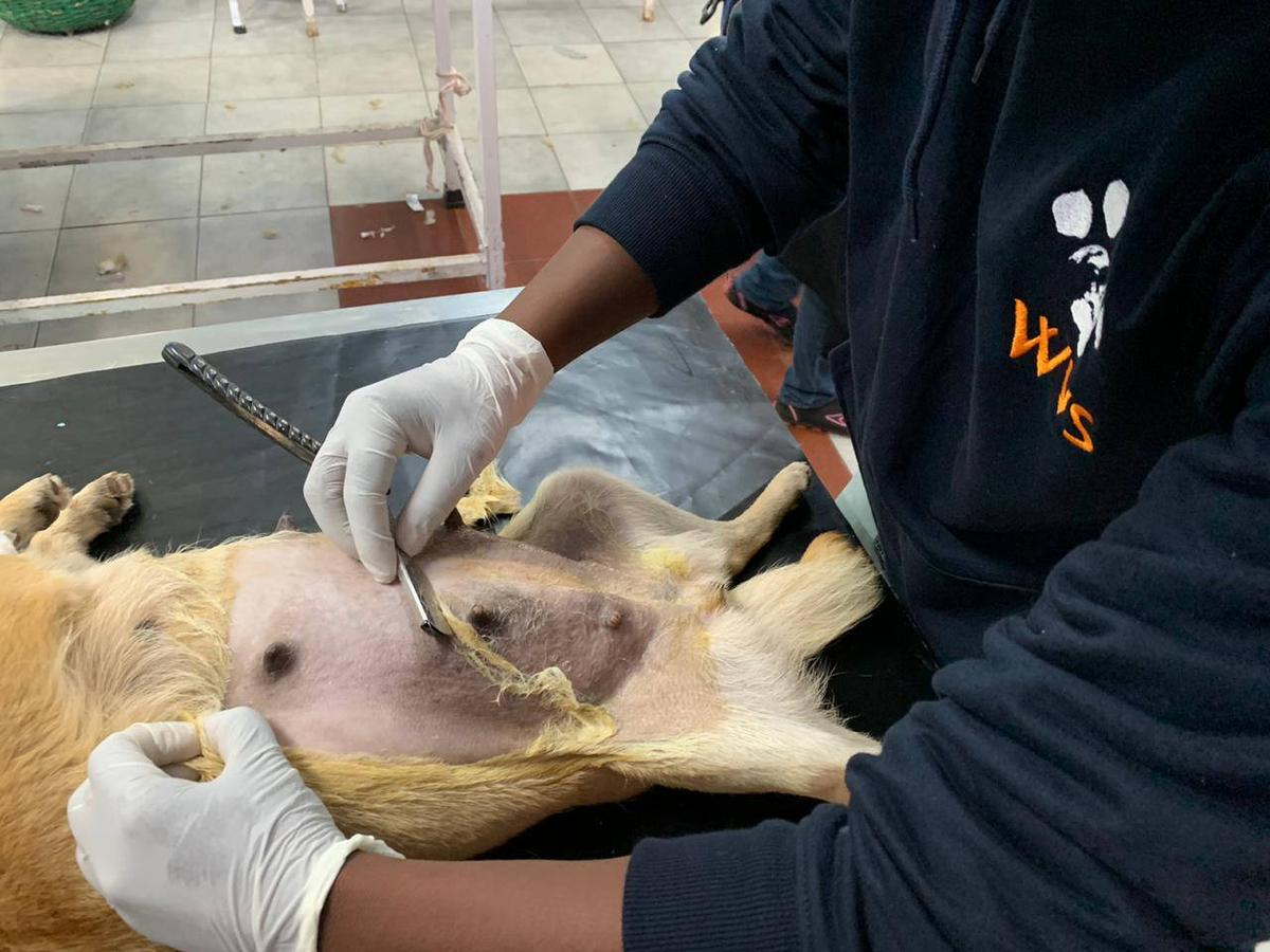
Pull. Always pull the blade towards you (do not push the blade away from you), with the blade perpendicular to the direction of shaving and at an acute angle to the skin as shown in Figure 2.
With the growth. First shave in the same direction as hair growth to remove the majority of the hair. Skill is needed to gauge the degree of pressure to apply to the blade along with direction and angle.
Final shave. Finally carefully shave in the opposite direction of the hair growth to achieve a close shave, however particular care must be taken not to damage the skin whilst doing so.
Surgical site margins
The area that has hair removed should be large enough to enable a skin incision to be enlarged, if necessary, or to prevent contamination by hair if the drapes move slightly. A general guideline is to remove around 15-20cm either side of the proposed incision site.
Bitch spay
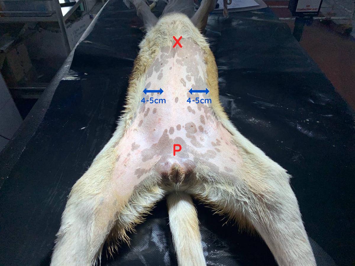
The clipped area should extend from the cranial margin (the xiphoid process), to the caudal margin (brim of the pubis). The lateral margins should extend 4-5 cm either side of the teats (depending on the size of your patient) (Figure 3).
For abdominal surgery always shave more than is required for a routine operation. In the event of surgical complications such as haemorrhage, it must be possible to safely extend the incision cranially and caudally whilst maintaining a sterile field - if you have not clipped enough this will cause a considerable difficulties for the surgeon and may reduce the chances of successful resolution for the patient.
Dog castrate
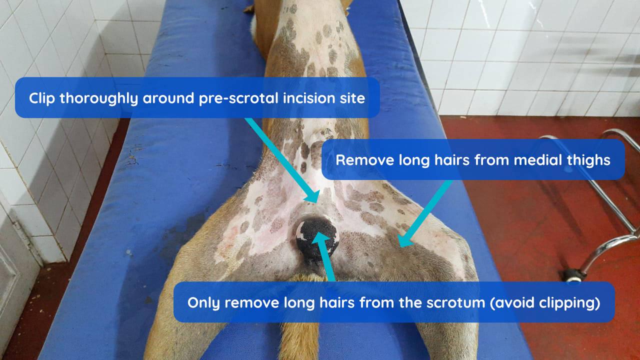
The prescrotal and prepuce areas should be clipped thoroughly, such that the incision can be extended if necessary. Avoid shaving the scrotal sac and inner thighs if possible, as the skin here is delicate and sensitive, however long hair should be removed to ensure that it does not interfere with the surgical site (Figure 4).
Hair and debris should be removed immediately from the clipped area and the preparation table using a vacuum or manual removal prior to beginning the surgical scrub.