

Following lavage, the next step is to debride necrotic (dead and dying) tissue. Necrotic tissue serves as a nidus (source) of infection, preventing the wound from progressing from the inflammatory phase to the proliferative phase of healing. Debridement is essential for any wound treatment, especially if primary closure is planned, to reduce the risk of wound breakdown and infection.
Ensure the fur around the wound is clipped prior to debridement. This ensures we can visualise the entire wound and easily see the borders between necrotic and healthy tissue.
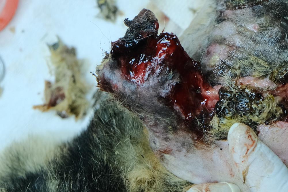
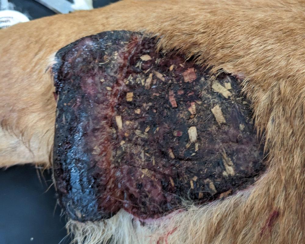
Recognising necrotic tissue
Necrotic tissue may appear black or green and stringy (similar to pus).
Methods of debridement
Different stages of wound healing require different debridement methods. Initially, more aggressive methods (surgical and mechanical) are necessary, but as healing progresses, selective debridement (autolytic, enzymatic, biotherapeutic) becomes more appropriate. Check for and debride necrotic tissue each time dressings and bandages are changed.
Surgical debridement
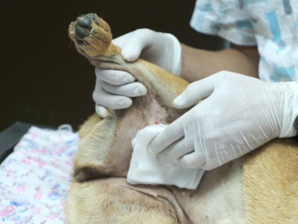
Surgical debridement is the gold standard to remove large amounts of necrotic tissue. Sharp dissection with a scalpel or scissors is used to remove all necrotic and devitalised tissue, with a small amount of healthy tissue being removed in the process. General anaesthesia or sedation and analgesia are used. After initial clipping and lavage, the skin around the wound is prepared aseptically using chlorhexidine or iodine solution. The vet can then begin the surgical debridement.

Instruments:
- Scalpel blades (No. 11 and 15), rat-toothed and atraumatic forceps.
Techniques:
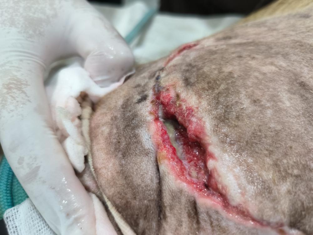
- Layered debridement. Begin by excising superficial necrotic tissue and removing debris, then move to deeper tissues. Look for the “line of demarcation” to distinguish necrotic from healthy tissue. Cut tissue back so normal-coloured tissue remains, and stop when bleeding occurs. Tissue that bleeds is healthy, and indicates debridement is sufficient. But don’t debride excessively.
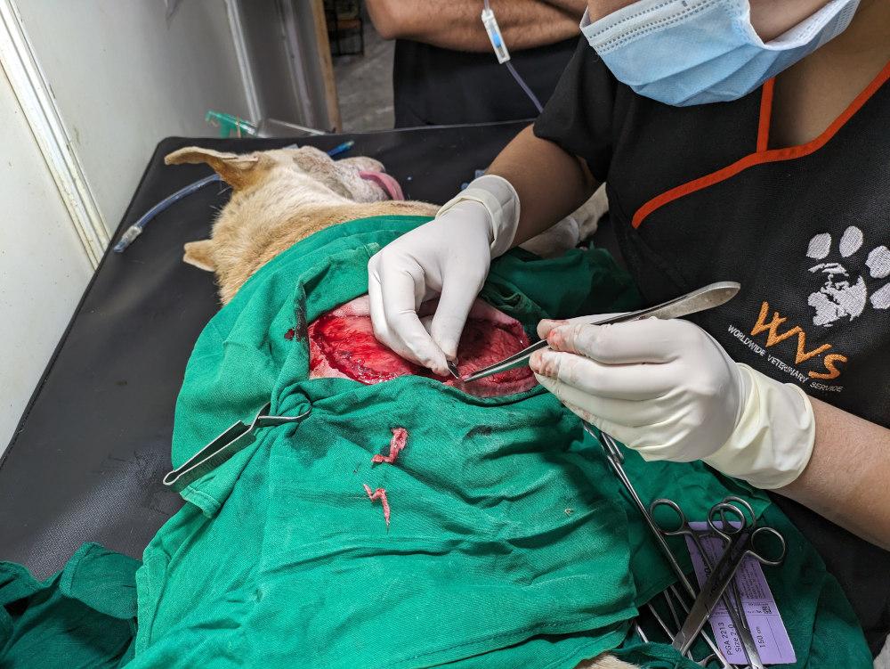
- En-bloc debridement. Remove the entire affected necrotic tissue, leaving a border of healthy tissue. The remaining healthy tissue may be sutured closed, allowing primary closure of the wound.
Mechanical debridement
Mechanical debridement uses dressings and bandages to remove smaller debris and contaminants not removed via surgical debridement.
Techniques:
- Wet-to-dry dressing. These are a controversial dressing choice. Indicated for wounds significant contamination and visible debris. Wet gauze is placed over the wound, bandaged, and allowed to dry, adhering to debris and necrotic tissue which is then removed with the dressing. This method damages underlying healthy and healing tissue and is very painful upon removal. They often slow down healing, but they can be a useful technique in some challenging situations with limited equipment.
Selective debridement
Selective debridement only targets necrotic tissue, preserving healthy tissue. Suitable for use after initial surgical debridement, as selective debridement works too slowly for primary debridement.
Techniques:
- Autolytic debridement. Uses the body’s own enzymes (collagenase, elastase) and phagocytes to destroy necrotic tissue. This slow process is enhanced by maintaining a moist wound environment, with moisture-retentive dressings (MRDs) such as calcium alginate, hydrogels, hydrocolloid and polyurethane foam. Honey is also an effective dressing choice, due to its antibacterial properties and promotion of autolytic debridement.
- Enzymatic debridement. Utilises dressings containing enzymes that break down necrotic tissue. These are less commonly available but can be effective.
- Biotherapeutic debridement. Involves the use of medical-grade maggots (disinfected Lucilia sericata larvae) to consume necrotic tissue. These maggots selectively eat necrotic tissue, also excreting enzymes that prevent bacterial growth, but leave healthy tissue in place.