

After manual debridement, a dressing is placed in contact with the wound and then secured with a bandage (see next article). In the inflammatory phase, the goal is to eliminate debris and bacterial contamination and encourage debridement. In the proliferative phase, however, the aim is to provide a moist wound environment that enhances the body’s healing processes. Appropriate dressings at each stage encourage healing to progress, from the inflammatory phase to the proliferative phase, and then into the remodelling phase.
Inflammatory phase wound dressings
During the inflammatory phase (typically lasting 1-5 days, ending when healthy granulation tissue is established), dressings should encourage additional debridement and remove bacteria and debris to prevent infection. Common choices are hyperosmotic dressings (honey, sugar), or antibacterial dressing (silver sulfadiazine). In exceptional circumstances, wet-to-dry (or dry-to-dry) dressings may be used, though these can damage a great deal of healing tissue and be very painful to remove.
Hyperosmotic dressings
Hyperosmotic dressings. dressings draw fluid out of wounds, carrying debris and bacteria out and onto the dressing, destroying bacteria cell walls through hyperosmolarity, thus reducing the infection risk.
- Honey dressing. It is cost-effective, readily available and has great antibacterial properties due to its hyperosmolarity, hydrogen peroxidase content, acidic pH, and antioxidants. These produce a great environment for wound healing, making honey the dressing of choice for most wounds in this phase. Sterile manuka honey is the best option, but in resource-limited environments, any raw honey may be used due to its price and availability. Apply honey to cover the wound, place a primary dressing layer over the top and secure with a bandage (secondary +/- tertiary layer). Replace the dressing daily initially, then every 2-3 days until healthy granulation tissue appears and the wound becomes less exudative.
- Sugar dressing. Though less effective than honey (lacking its antibacterial properties), sugar’s osmotic action draws out bacteria and benefits most inflammatory wounds. Mix sugar with a small amount of saline to form a paste, place it on a dressing layer (e.g. sterile swabs), then cover the wound and secure it with a bandage.
Antibacterial dressings
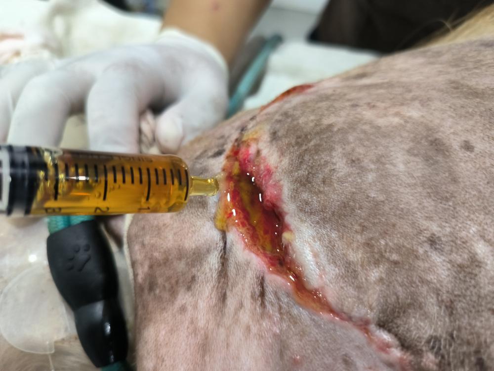
Antimicrobial dressings use a topical antibiotic paste applied within the wound, kept in place with a non-adherent dressing or clean swabs and a bandage, to directly attack bacteria.
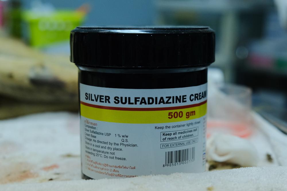
- Silver sulfadiazine. Silver is toxic to bacteria and yeasts, while sulfadiazine is a broad-spectrum antibiotic. This combination effectively kills bacteria within the wound, facilitating the progression of the healing phases. However, honey is generally preferred due to its reduced risk of antibiotic resistance and its additional healing benefits.
Adherent dressings
Adherent dressings adhere to the wound and cause mechanical debridement when removed, which can be useful for visibly dirty wounds. However, these may be painful and can damage healing tissue.
- Wet-to-dry dressings. This method is less preferred due to being very painful when removed and damaging a great deal of healing tissue. Better alternatives are usually available (honey dressings), however, wet-to-dry dressings may be used in heavily exudative wounds with lots of gross contamination (visibly dirty wounds) where appropriate pain relief is on board. Gauze swabs are soaked with sterile saline, placed on the wound, and secured with a bandage. The swabs dry and adhere to the wound, then are removed the next day to debride it.
- Dry-to-dry dressings. These use the same process as wet-to-dry dressings, but the gauze swabs are not made wet. Dry swabs are placed over the wound and secured with a bandage. Leave for 12-24 hours then remove to mechanically debride. These are used for slightly less exudative wounds, however, they are also painful dressings and are mostly avoided.
Wound dressings for the proliferative phase
When a wound progresses to the proliferative phase (when a healthy bed of granulation tissue has formed), our aim shifts to maintain a moist wound environment to facilitate natural healing. Due to a lower risk of infection, dressings may be changed less frequently. The proliferative phase lasts 4-21 days, depending on the wound.
Once a wound reaches the proliferative phase, we can consider closing it using delayed primary closure, so long as the wound can be closed without undue tension, and no infection or necrotic tissue is present. Closure may speed up the process of healing. Ongoing fluid accumulation in a closed wound can be removed by placing a drain.
Exogenous moisture dressings
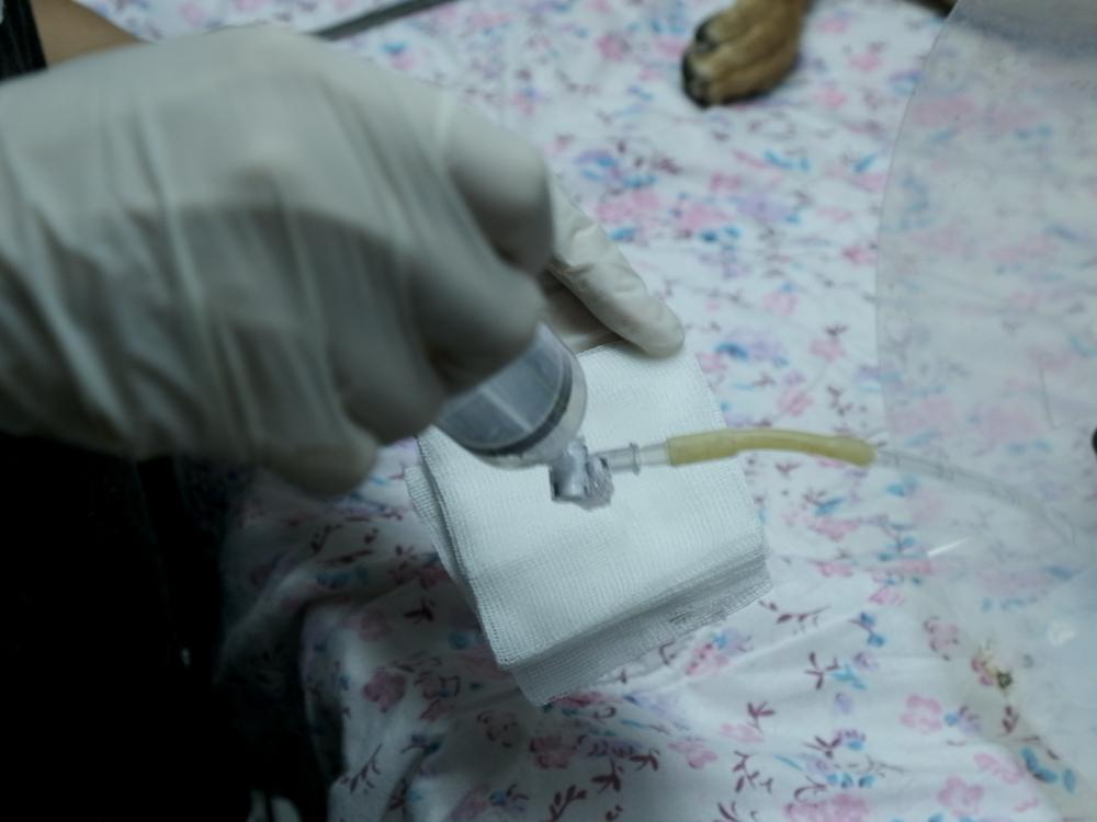
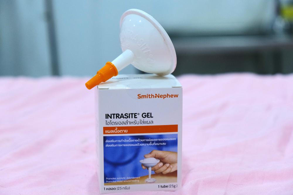
- Hydrogels (e.g., Intrasite). These add moisture to dry, non-exudative wounds in the proliferative phase. Apply the hydrogel to the wound, cover with a non-adherent dressing, and secure with a bandage.
Moisture retentive dressings
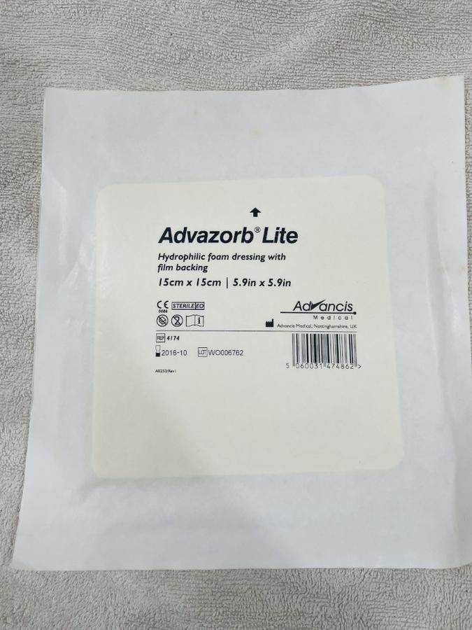
- Foam dressings, Alginate. Suitable for slightly exudative wounds, these dressings maintain a wound’s natural moisture. The dressing is placed with a bandage to secure it.
Non-adherent dressings
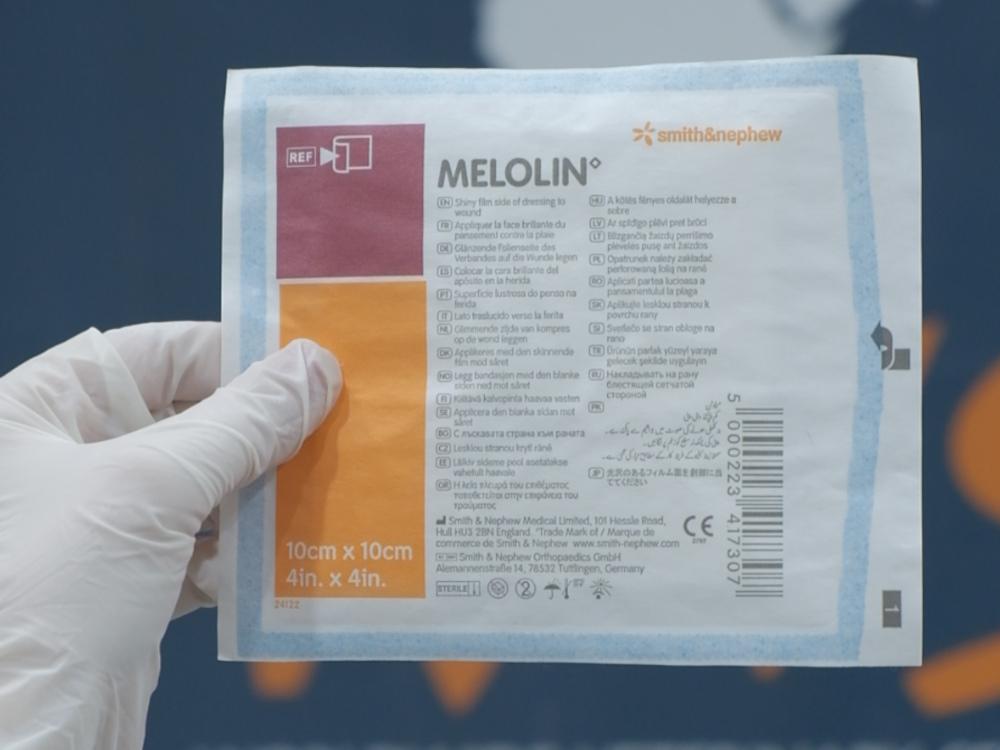
- Melolin, Telfa. Ideal for more exudative wounds. These dressings absorb excess moisture, without adhering to the wound, reducing trauma during dressing changes. Secure these with a bandage.
Silver sulfadiazine cream
Silver sulfadiazine cream is also used during the proliferative phase, it provides antibacterial action and maintains moisture in the wound (optimal healing environment). A non-adherent dressing is placed above and secured with a bandage.
Incorrect dressings
Using an incorrect dressing can impair healing, which may lead to complications. Below are some examples of the use of an incorrect dressing.
- Adherent dressings with granulated wounds. Using wet-to-dry or dry-to-dry dressings on wounds in the proliferative phase will cause trauma to the delicate granulation tissue, and delay healing. At this phase of healing, wet-to-dry dressings will not improve patient outcomes.
- Honey dressing with a granulated wound. Once in the proliferative phase, a moist environment is required to progress healing. Honey’s hyperosmotic action draws water out of the wound, meaning it would dry out granulation tissue and impede healing. As such honey shouldn’t be used at this point.
- Non-absorbent dressings on exudative wound. Applying dressings that don’t absorb moisture on a highly exudative wound means the surrounding tissues will be exposed to excessive moisture. This may lead to maceration (softening and breakdown) of the surrounding tissues, which increases the risk of infection and slows healing.
- Occlusive dressings on infected wounds. Occlusive dressings (like Vaseline) prevent air from getting to the wound and may encourage the growth of anaerobic bacteria within a wound.
- Overuse of antibacterial dressings (silver sulfadiazine). Especially in wounds which are not infected, this may lead to antibiotic resistance and their use isn’t necessary in non-infected wounds.
Conclusion
By selecting an appropriate dressing for each phase of wound healing, clinicians enhance the healing process, preventing infection and encouraging progression from inflammation, to proliferation and finally maturation. Next, we discuss various bandaging techniques to hold dressings in place.