

Patient preparation
Food. This should only be withheld if the patient is to be sedated/ anaesthetised, or if dysphagia or other gastrointestinal issues are being investigated, and abdominal radiographs are to be taken as well.
Care whilst positioning. Many animals undergoing thoracic radiographs are already in respiratory distress. Some positions (lateral) increase the stress on the already compromised respiratory system. Take care to minimise the amount of time patients spend in these positions, or avoid them entirely if they are not needed. Use positioning aids as needed, but try not to place them in a way that may increase respiratory distress (heavy sandbags over the trachea etc).
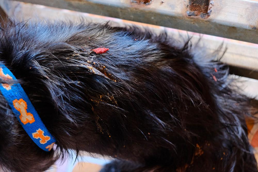
Clean coat. Dirt should be cleaned off the patient, and the coat should be dry, to minimise artefacts.
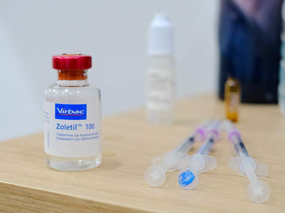
Sedation/ General anaesthesia. Decide whether the patient requires sedation or not. Ensure patients aren’t sedated with agents that may further compromise respiratory function. Sedation or general anaesthesia allows easier positioning, less patient stress, less motion blur, and manual lung inflation. However, general anaesthesia may cause the collapse of alveoli (reducing visibility), and increases risk in already sick patients. Manual inflation of the lungs (using a breathing-bag) may lead to over-inflation, which will make it harder to see some pathologies, like small nodules within alveoli.
Standard radiographic views
The standard views we may use for thoracic radiographs are:
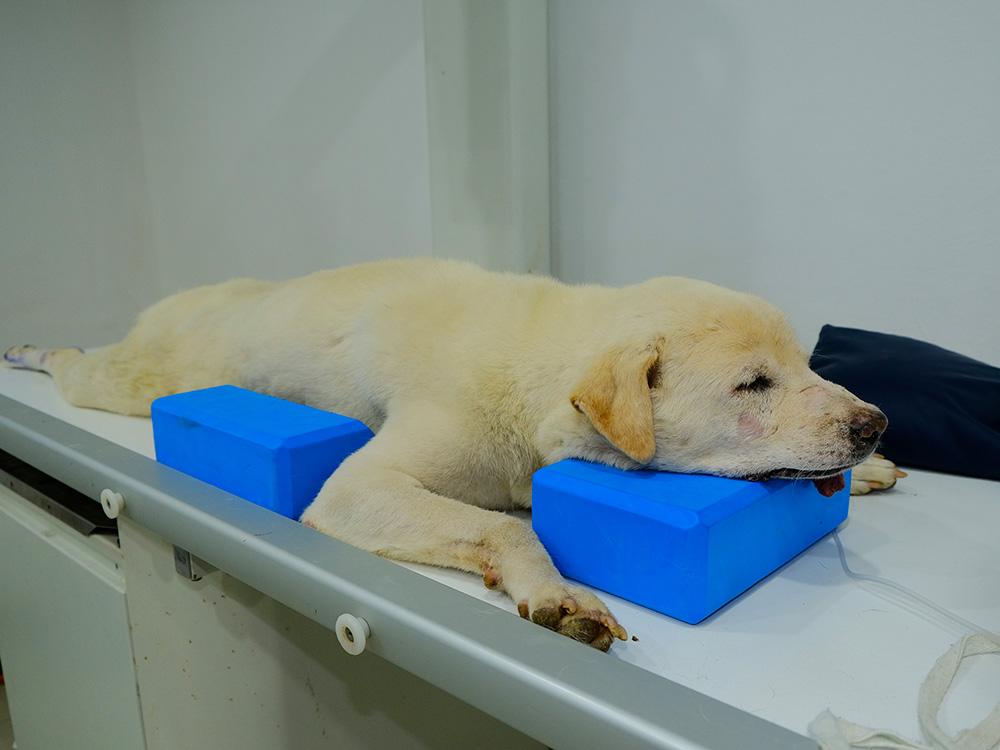
Dorsoventral. The patient lies with their sternum on the plate, legs outstretched. This view lets us lateralise a lung lesion and also gives information about the heart’s size and shape. It also leads to better inflation of the caudodorsal lung fields, improving visualisation.
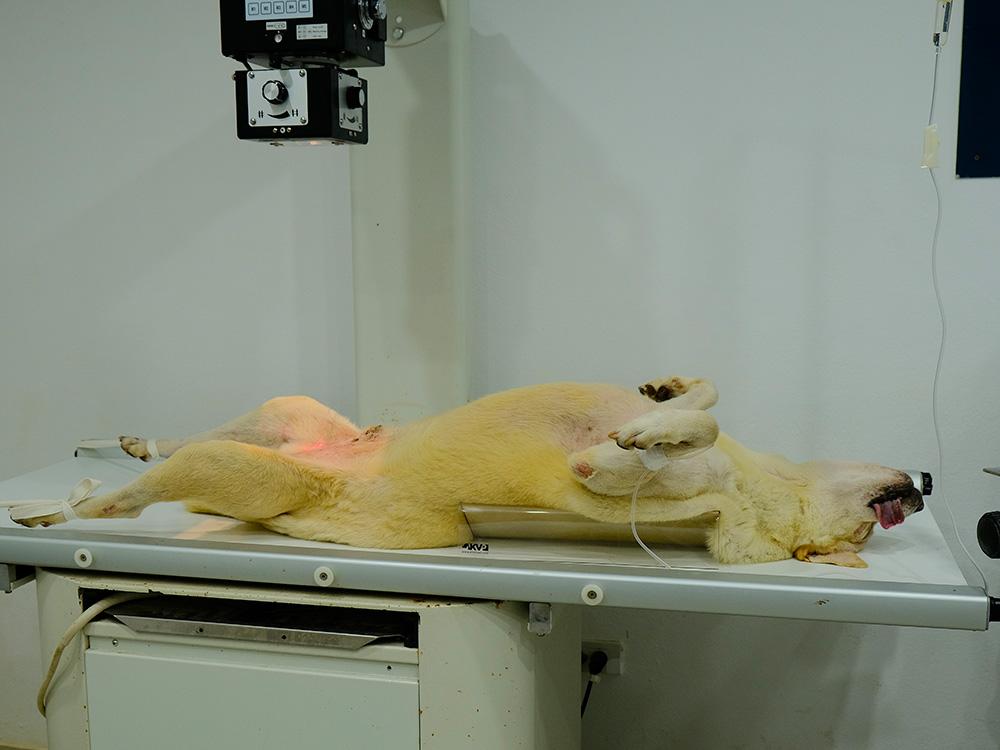
Ventrodorsal. The patient is lying on their back, legs in the air. This view allows better visualisation of the accessory lung lobe and the cranial ventral lung fields. However, it should not be performed if pleural fluid is suspected, or the patient is severely dyspnoeic.
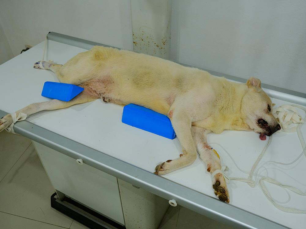
Right and left lateral. The patient is lying on their side. Minimise time spent in this position for dyspnoeic patients, as the position increases respiratory distress (more difficult for lungs to inflate). Always take both laterals when looking for subtle changes like metastasis in the lungs.
The minimum views required for identifying specific conditions are:
Cardiac conditions. Dorsoventral view and right lateral, allowing us to see the cardiac silhouette.
Lung conditions. Ventrodorsal view and right lateral, showing us the lung fields.
Lung metastasis. Ventrodorsal view, left AND right lateral so we don’t miss possible metastases on the opposite side.
Exposure considerations
High kV, low mAs. The thorax is highly mobile due to the lungs moving with breathing, so there is a greater risk of motion blur. To reduce this risk, we use a shorter exposure time and thus low mAs. The different tissues in the thorax vary greatly in radiodensity (bone, gas, soft tissues, liquid), so we need a high penetration which is achieved with a high kV. The variety of tissues in the thorax also means a good natural contrast, so we don’t need to increase mAs to increase it like we would in the abdomen, where the different organs are more homogenous.
Inspiratory view. This ensures the lungs are full of air and enlarged, which lets us see them in greater detail. We may do expiratory views to detect bullae, trapped air with feline asthma, or a pneumothorax.