

The clinician should only perform thoracic radiography when it is appropriate. In some cases, ultrasound may be a more beneficial imaging technique.
Common indications for thoracic radiography include:
Coughing. A patient with severe acute or chronic coughing may benefit from thoracic radiography. Possible underlying causes include primary pulmonary disease (inflammatory), left-sided congestive heart failure, primary or secondary neoplasia, foreign body inhalation (grass seed), or lungworm.
Dyspnoea (difficulty breathing). Cats commonly present with dyspnoea. Whilst this may be due to severe stress following a traumatic incident, radiographs are important as it may also be due to pleural effusion secondary to trauma (road traffic accident). Any insult to the airways may cause dyspnoea, including obstruction, pulmonary disease, or other pleural disorders. Many causes of dyspnoea can be diagnosed through radiography.

Cardiovascular disease. An ultrasound is needed to properly assess cardiac function. Coughing may be secondary to cardiovascular disease, however, an enlarged cardiac silhouette paired with normal lungs may indicate that the heart is the primary source of the disease. In this case, a cardiac ultrasound may then be performed to better assess its function.
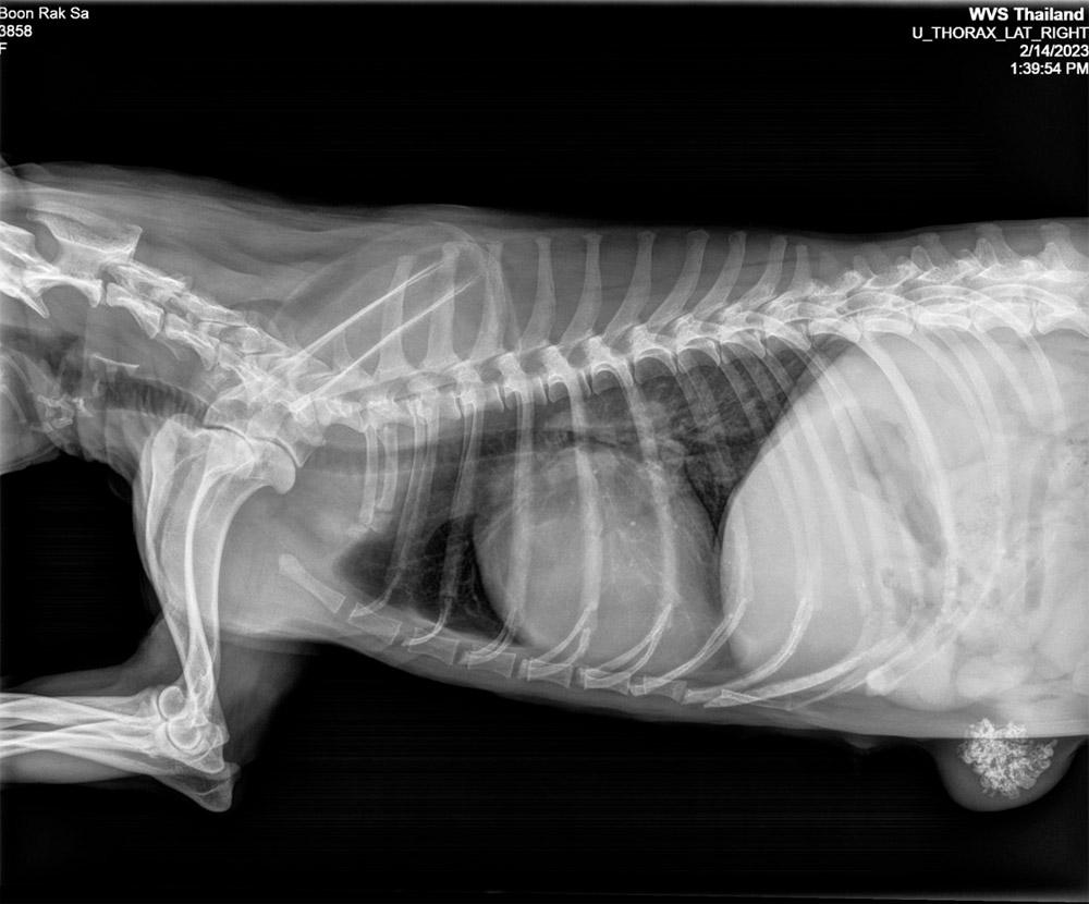
Trauma. Following RTAs, thoracic radiographs let us assess for damage within the thorax. This is done most often in cats. Common findings following trauma are diaphragmatic rupture and herniation, pleural effusion, pneumothorax, haemothorax, and possibly pyothorax in the days following the trauma. Thoracocentesis or surgery may be necessary for treatment.
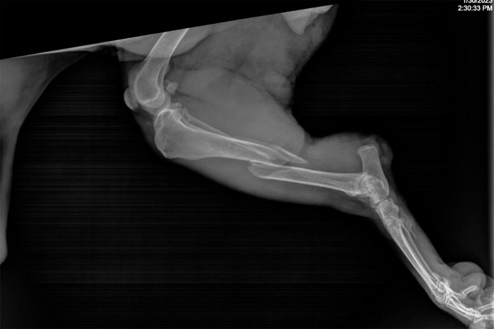
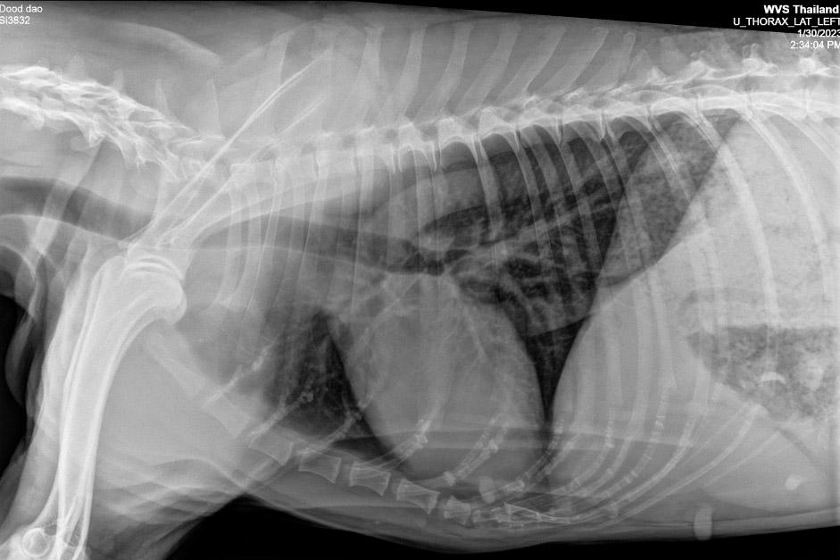
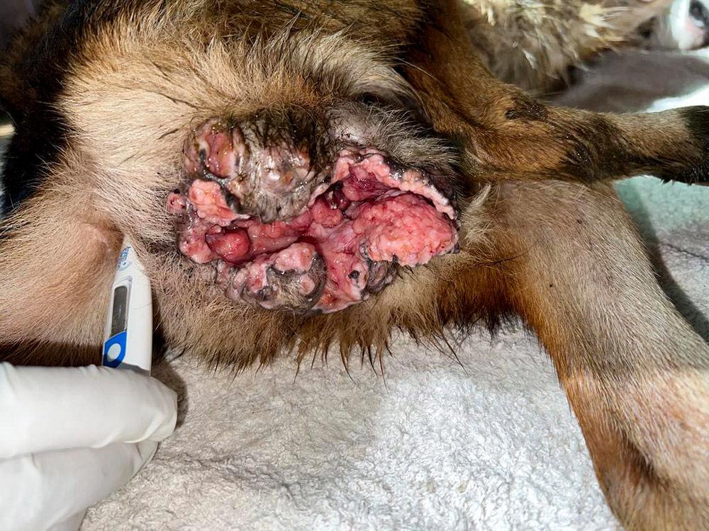
Neoplasia. We may radiograph the thorax due to a suspected primary neoplasia (following coughing, reduced energy in the older patient etc), but we might also radiograph to determine whether secondary metastases have occurred. The lungs are a very common site of metastases for many tumours, and detecting these can give us an indication of the animal’s prognosis. In osteosarcomas in the dog, 80% will die due to secondary lung metastasis (Szewczyk et al., 2015).
Dysphagia (difficulty swallowing). Thoracic radiographs may allow us to detect the aetiology of regurgitation disorders. Lateral radiographs may diagnose megaoesophagus, some congenital issues, and foreign bodies.