

1. Select exposure settings on the x-ray machine, and place the grid on the plate (where the depth is over 10cm).
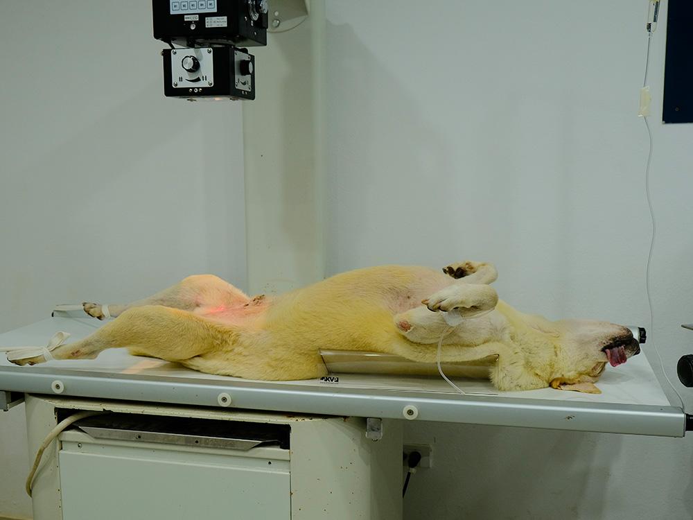
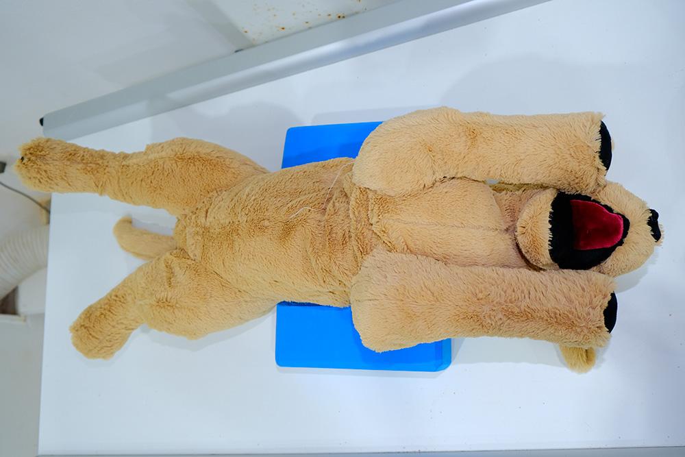
2. Position sedated patient in dorsal recumbency (spine to the plate with legs in the air and hindlimbs frog-legged). The patient may be placed in a positioning trough.
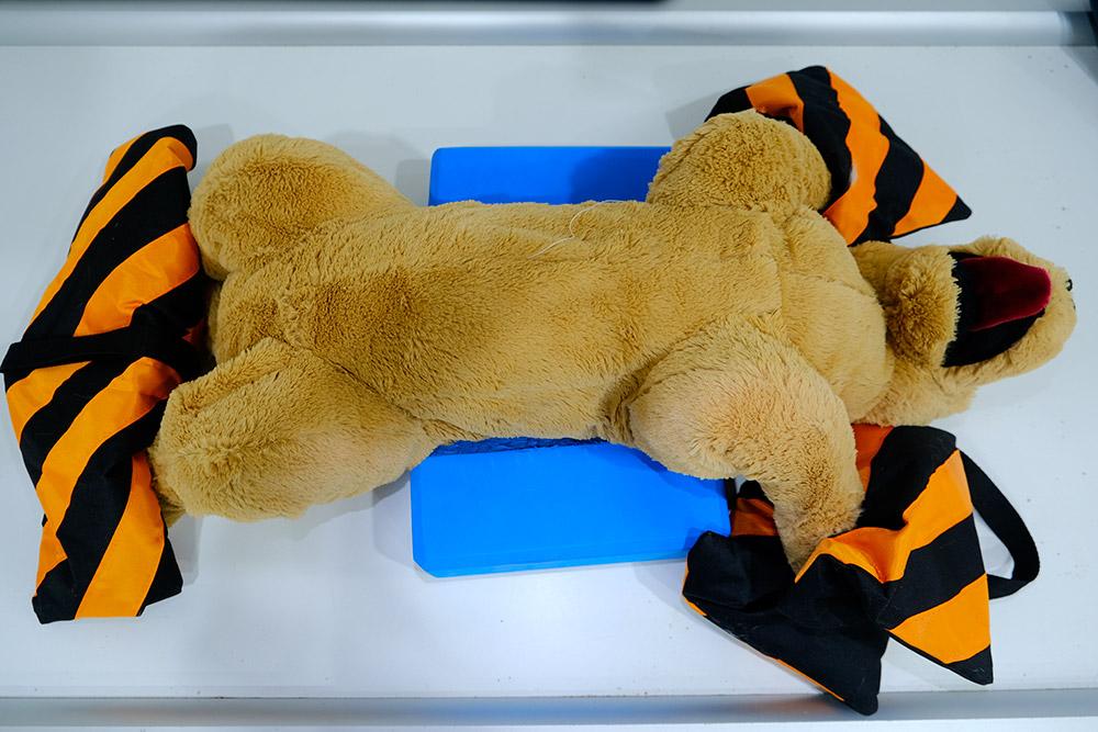
3. If no positioning trough is used, place foam wedges on either side of the patient's chest to minimise rotation.
4. Place sandbags over the forelimbs and hindlimbs to hold the patient in the desired position.
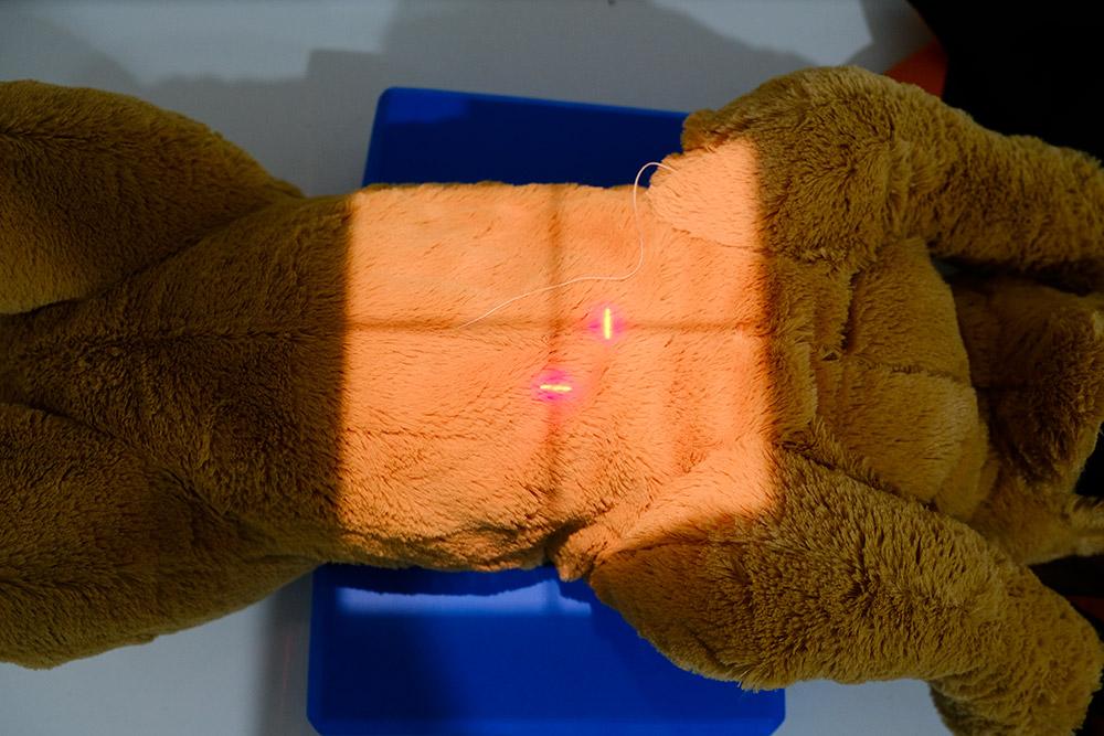
5. Centre the primary beam over the mid-thorax.
6. Collimate the primary beam cranially to the thoracic inlet, caudally to the last rib, and laterally to include both skin edges.
7. Place the right/left marker within the beam's collimation on the corresponding side.
8. Take the radiograph.