

A microscopic “wet sediment” examination can be performed cheaply in-house, for rapid results, detecting the abnormal presence of cells (leucocytes, erythrocytes, epithelial cells), bacteria, casts and various crystals. With practice, vets can become quick at accurately identifying and differentiating these findings. Determining crystal types is especially important to diagnose and treat urolithiasis.
How to prepare the sample
- Obtain a fresh urine sample. If using a refrigerated sample, warm it to room temperature first and gently mix it. Use a standard volume (usually 5ml*).
- Transfer it to a cone-shaped centrifuge container.
- Centrifuge at 1000-1500 rpm for 5 minutes. Lower speeds help to preserve the sediment without damaging it.
- Discard the supernatant (liquid on top of sediment). Using a pipette or by decanting, remove the majority of the supernatant leaving approximately 0.5ml.
- Optional: add a stain (such as Sedi-stain) to the sample, otherwise continue without staining.
- Flick the tube gently to re-suspend sediment in the remaining supernatant.
- Pipette 1-2 drops of urine onto a microscope slide.
- Place a coverslip over it ready for examination.
*The concentration of solids in a urine sample is volume dependent; therefore, use the same volume of urine each time to ensure an accurate assessment.
Examining the slide
- Lower the microscope condenser (to increase contrast), especially if the sample is unstained.
- Place the slide under the x10 lens, and inspect the entire slide for crystals.
- Then, use the x 40 lens for casts, smaller crystals, cells and bacteria. This magnification is considered ‘high power’ and the numbers of cells or crystals should be counted to evaluate the number per high power field. Examine 10 fields of view, and calculate the average values.
To help confirm the presence of bacteria, a x100 oil immersion lens may be used. Stains added to the wet preparation (e.g. Sedi-stain) or to an air-dried sample (e.g. Diff-quik, Giemsa, Gram) can also be useful.
Possible findings in sediment exam
Cells
Erythrocytes. Also known as haematuria. Seeing a few erythrocytes may be normal (for cystocentesis samples), however large numbers suggest urinary tract infection, inflammation or iatrogenic trauma (poorly executed cystocentesis or catheterisation).
<5 erythrocytes per high powered field (HPF) can be found in the urine of healthy dogs and cats (1).
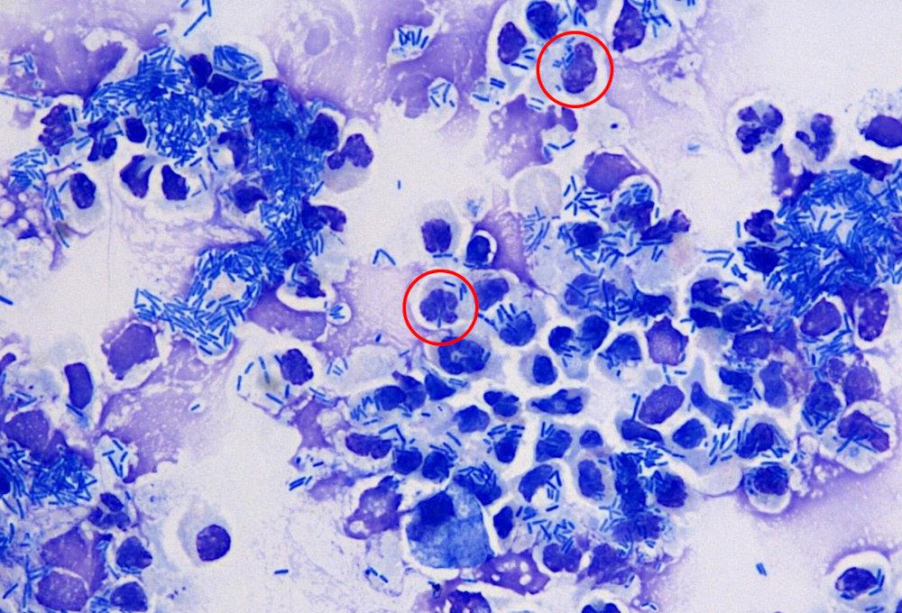
Leucocytes. Also called pyuria. Neutrophils are most common. Normally these would not be present, but numbers increase with urinary and genital tract infection and inflammation.
<5 leucocytes per high powered field is normal for healthy dogs and cats (1).
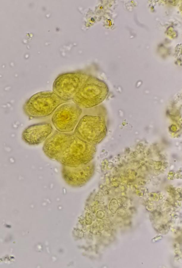
Epithelial cells. These may be shed from the kidneys, bladder, urethra, vagina, vulva or prepuce. Low numbers, no more than two epithelial cells under a x10 (low power) view, are likely to be normal. However, increased numbers may be associated with inflammation, hyperplasia or neoplasia. It can be challenging to differentiate between the types of epithelium (e.g. squamous from lower urinary tract; transitional from bladder, ureter or renal pelvis).
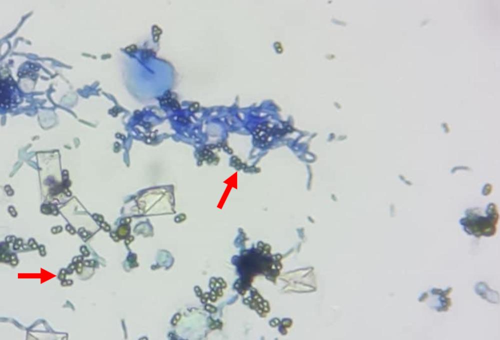
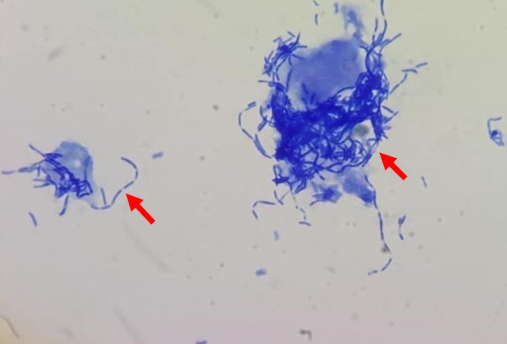
Bacteria. Interpret the presence of bacteria in the context of sample collection and clinical signs. Their presence is most likely to be clinically significant in cystocentesis samples where contamination is unlikely to occur, whereas free catch samples can contain bacteria from the environment. The absence of bacteria in a sediment sample does not rule out infection. Bacteria will appear as small rounded cocci, or more elongated rods - and may be in clusters or chains.
Casts
Casts are formed in the lumen of the renal tubules. They are composed of protein and may also incorporate cells and other materials. In general, they are absent in the urine of a healthy animal. It is important to note that casts are shed intermittently and so their absence does not rule out disease. The type of cast is less important for interpretation, and rather their presence, especially in high numbers, is a cause for concern, indicating disease in the renal tubules. They may be recognised under low power with a distinctive cylindrical shape.
Hyaline casts. Protein precipitates which may occur due to exercise or kidney tubular disease. They appear cylindrical and colourless.
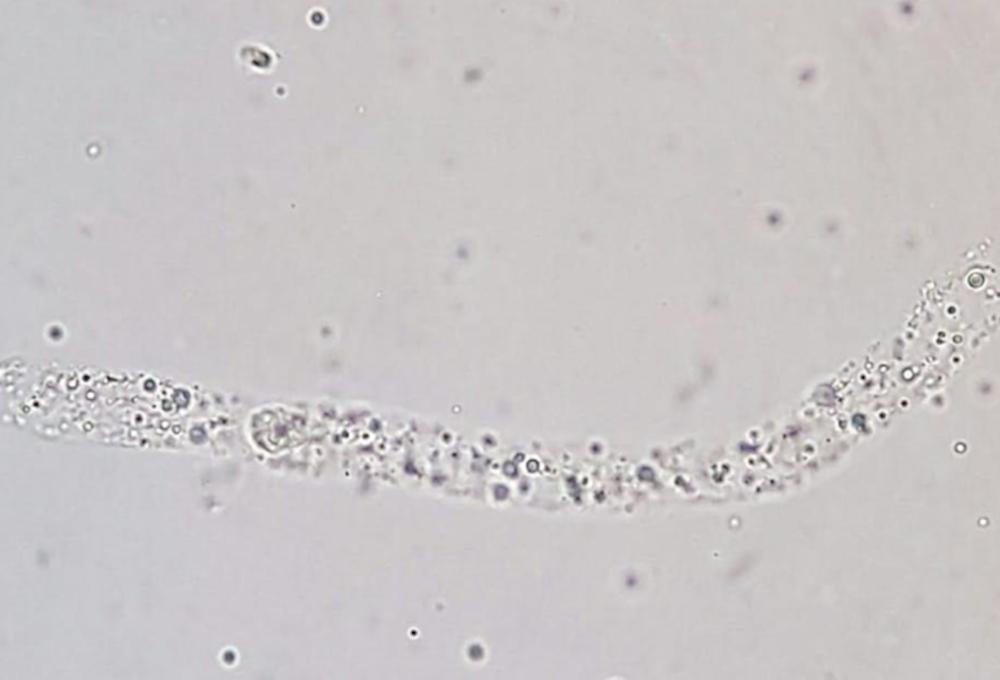
Cellular casts. These may contain erythrocytes, leucocytes or epithelial cells. These are always abnormal. Erythrocyte casts appear due to renal haemorrhage, and leucocyte casts due to renal inflammation.
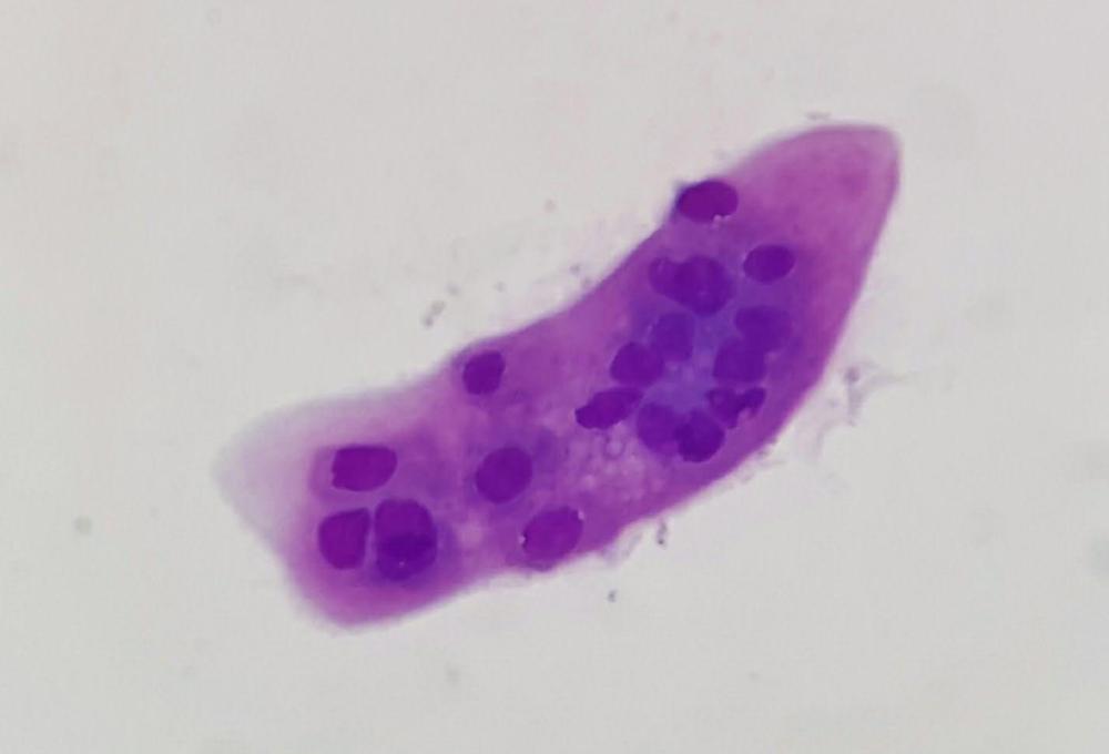
Granular casts. These represent degenerated epithelial and white blood cell casts, and occur alongside significant disease of the kidneys. These may deteriorate into waxy casts, suggesting chronic renal disease.
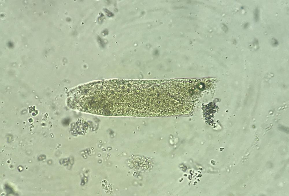
Note: finding <2 granular or hyaline casts per low powered field may be normal, but higher numbers, and any number of cellular casts should always be considered abnormal.
Crystals
Remember, a fresh sample is vital as crystals may dissolve or form over time, including with temperature change. Some crystals may be seen in healthy patients, so we should consider their significance alongside clinical signs. Crystals may indicate urinary tract infection, but also the type of urolith present in animals with bladder stones or blockages.
Some of the more commonly seen crystals are described below.
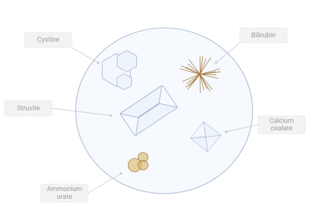
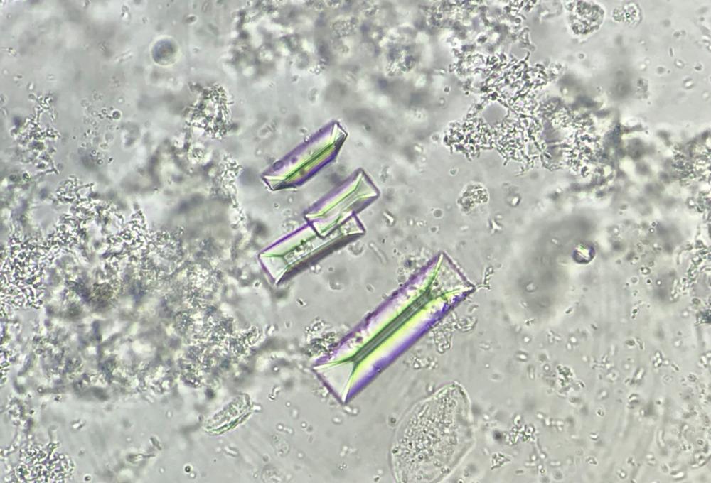
Struvite (magnesium ammonium phosphate).A common type of crystal in cats and dogs, and may be an incidental finding (normal in low numbers). They can be seen at low magnifications, and are most common in concentrated and alkaline urine. They may be present with a urinary tract infection, particularly in dogs. They have a distinctive appearance as colourless, three-dimensional prism-like crystals, sometimes described as ‘coffin shaped’.
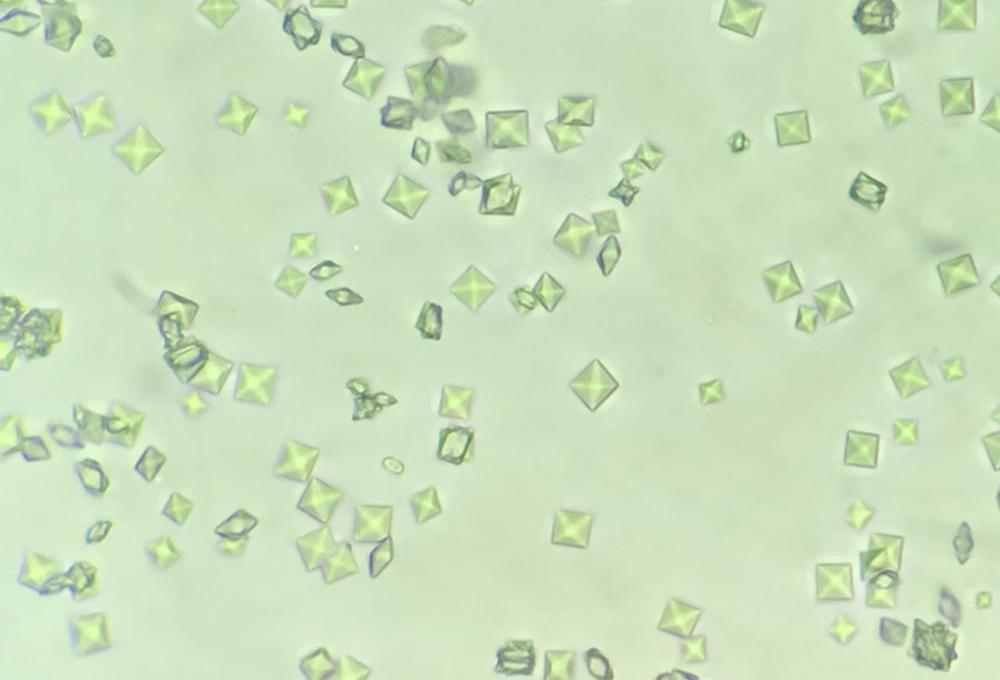
Calcium oxalate (dihydrate). Occur commonly in cats and dogs but also may be incidental findings. These crystals are common in acidic urine. They have a distinctive square shape, with a cross, and vary greatly in size. The calcium oxalate monohydrate variation looks slightly different, and presents with ethylene glycol toxicity (or hypercalcaemia) (see below).
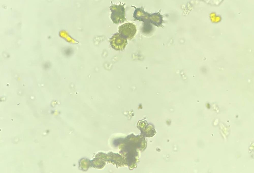
Ammonium urates. Relatively rare, ammonium urate and biurate are associated with liver disease (i.e. portosystemic shunts). Some breeds are predisposed to forming these due to a genetic mutation, e.g. dalmatians. These appear as yellow brown spheres, amorphous structures or diamond shapes.
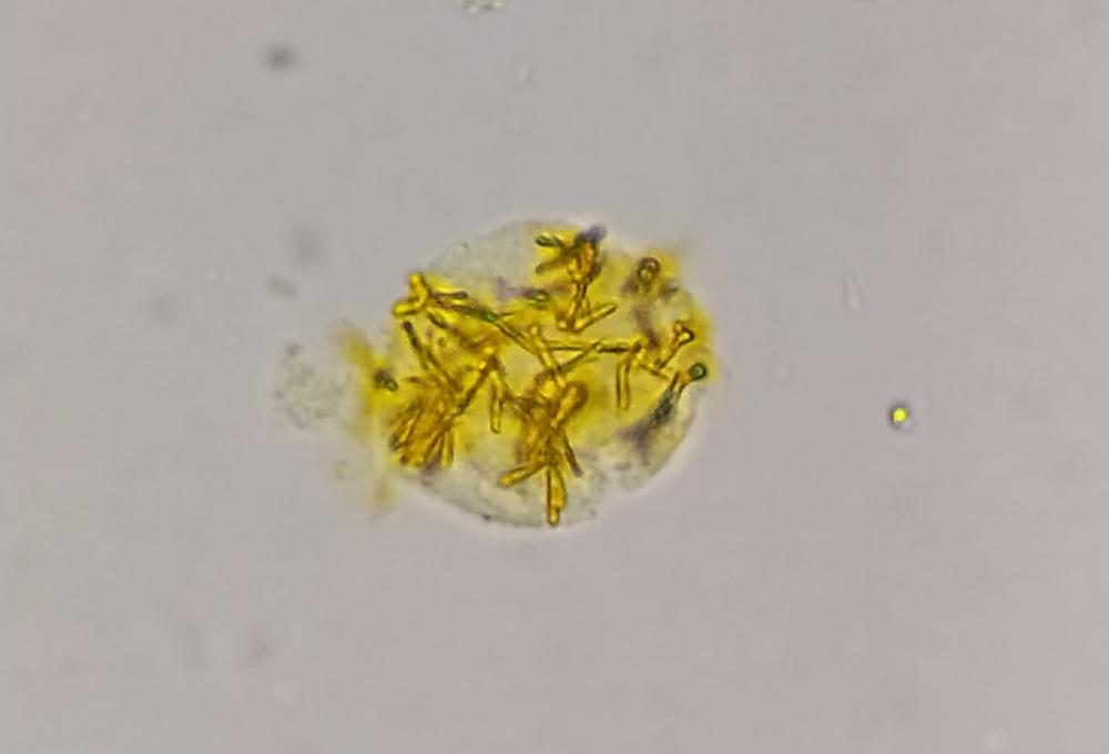
Bilirubin crystals. Can be a normal finding in healthy dogs producing concentrated urine, but are always considered abnormal in cats. Vary in size, usually small and needle-like yellow to yellow-brown colour. Bilirubinuria can be associated with liver disease, or the breakdown of red blood cells.
Amorphous material
These are aggregates of small crystals or granular material that do not have a defined shape and are often composed of phosphates, urates or xanthine. Sometimes they be similar in appearance to bacterial cocci and the use of a stain such as gram can help differentiate them. Finding these amorphous crystals does not lead to a specific diagnosis.
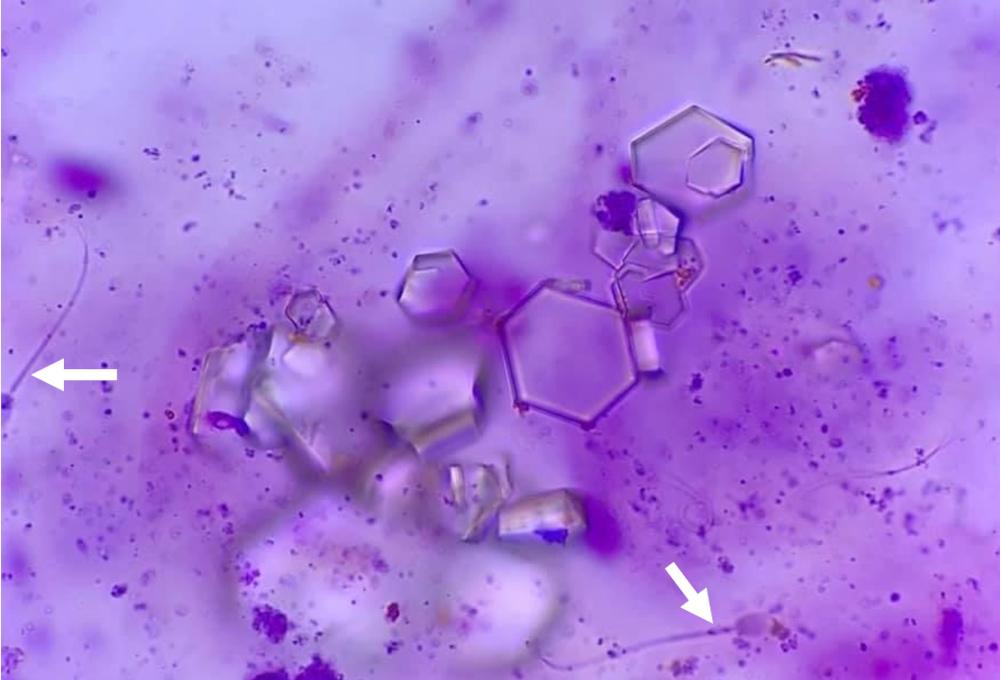
Cysteine. Considered pathological in cats and dogs. Appearance is hexagonal, flat and colourless. Associated with a tubular transport defect.
Calcium phosphate.
These are uncommonly found in neutral to alkaline pH. They are often small prisms or blunt-ended needles and can be arranged in a rosette pattern or amorphous.
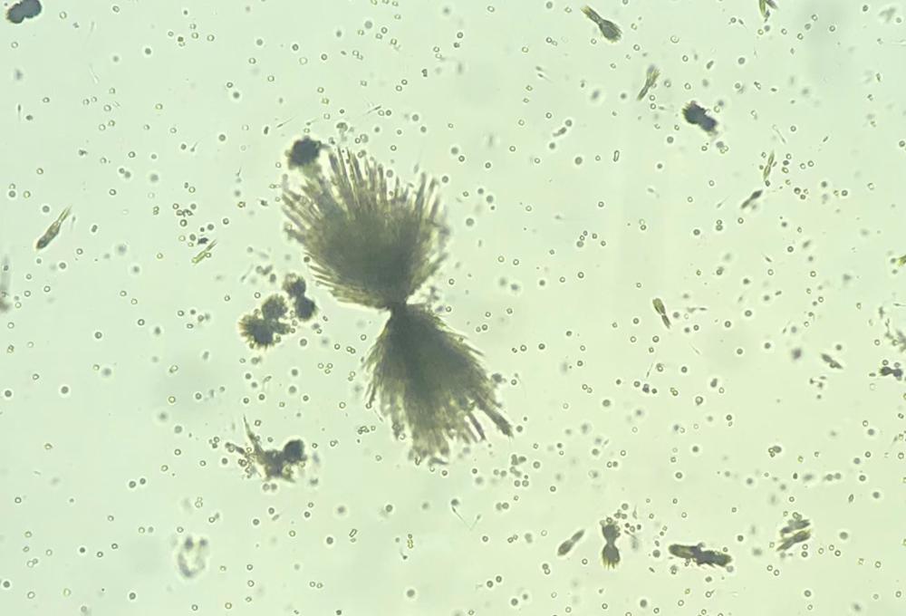
Calcium oxalate monohydrate.
These are uncommonly seen in healthy dogs and cats but can be associated with diseases associated with hypercalcaemia and hypercalciuria, such as lymphoma, or specifically after ingestion of ethylene glycol (anti-freeze solution). They have a characteristic “picket fence” appearance (straight sides with pointed ends) as well as more of an oval or dumb-bell shaped.
Sulphonamides. These are found in animals being treated with sulphonamide antibiotics. They are often needle shaped, dark and may appear in a fan shape.
Other
Sperm. Occasionally seen in healthy intact males (see Figure 8).
Fat droplets. Appear as spherical bodies, and may be due to sampling contaminants. They will appear on a higher plane of focus than cells.
Mucus. Not clinically significant and are secretions from the genital tract, appear as thread-like structures.