

Key Points
- The repair stage regenerates damaged tissueand restores structural integrity.
- It begins 3-5 days after the wound occurs (overlapping with the end of the inflammatory stage) and can continue for several weeks.
- The presence of granulation tissue is a hallmark of repair, and it has a moist, pink appearance.
- Epithelialisation restores the skin’s outer layers, and contraction of the wound brings the edges closer together.

Formation of granulation tissue
At around 3-5 days after initial injury, fibroblasts migrate into the damaged tissue and proliferate. Activity is well-established by 7 days. Depending on the size of wound, this fibroblast activity may continue for several weeks.
Fibroblasts synthesise collagen and other materials which convert the provisional extra cellular matrix (which is plugging the wound), into connective tissue, i.e. granulation tissue. Collagen provides structural support, and a foundation for regenerating tissues to build on. New capillaries also grow into the wound, delivering oxygen and nutrients, to support the healing tissue. This is called angiogenesis.
Over time, the wound is filled with a new, durable connective tissue. The fibroblasts begin to slow down their collagen synthesis. The new capillaries start to regress as the tissue heals, and the demand for blood supply reduces. This is why early granulation tissue looks darker pink/red, but in the later stages becomes paler.
Healthy granulation tissue will be pink, moist and homogeneous. A well-developed granulation bed is a good sign, and shows the wound is progressing towards closure.

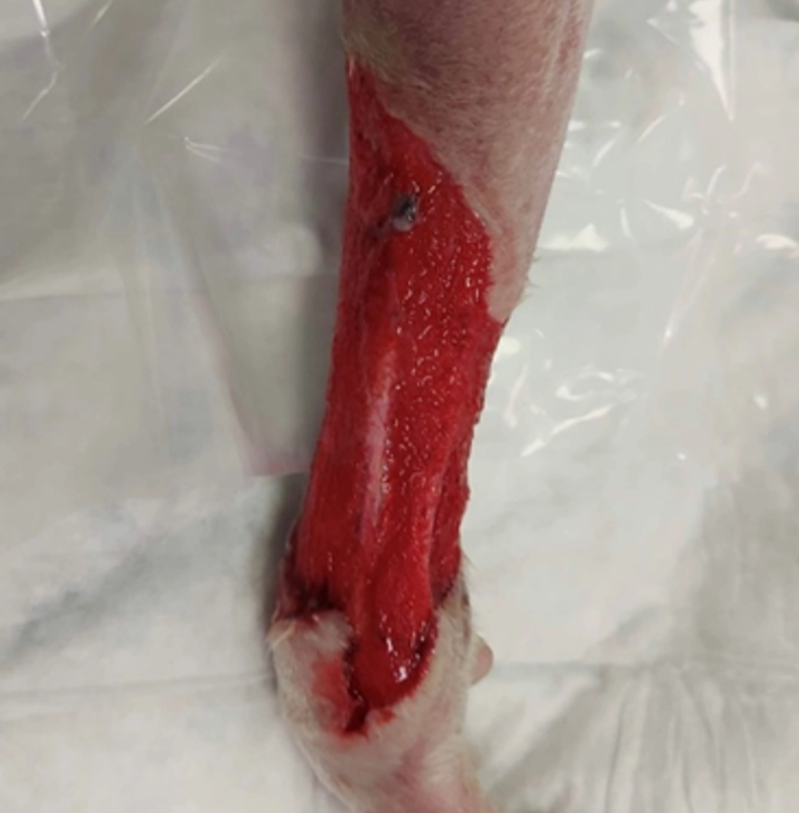
Figure 1 and 2 — Granulation tissue’s pink colour comes from the production of new capillaries (angiogenesis)
Contraction and epithelialisation
After granulation tissue is formed, the skin’s epithelium is regenerated to cover the wound. New epithelium starts to appear at the wound edges at around 4-5 days after injury, and continues until the wound is covered. Keratinocytes migrate from the wound edges, proliferating and form a new layer of skin. With time, the epidermis regains its stratified layers, and repigmentation can start 1-2 weeks after injury (but may take several months).
Meanwhile, specialised cells called myofibroblasts (a type of fibroblast containing smooth muscle proteins) infiltrate the wound. They work to contract the extra cellular matrix and granulation tissue, bringing the edges of the wound closer together. The surrounding skin stretches to accommodate this, and the wound can take on a stellate appearance (star shaped). Contraction reduces the size of the defect, and reduces the amount of scar tissue. Wound contraction may be visible from as early as 5-9 days after initial injury. In wounds healing by secondary intention, much of the process is achieved by contraction.
Contraction stops if there is too much tension on surrounding tissue. There may be occasions where the edges do not meet, and granulation tissue remains in the middle of the wound. In these cases, epithelialisation is often able to cover this remaining defect. However, epithelialisation may be incomplete in very large wounds, and in some cases an area of granulation tissue may remain, or only be covered by a thin layer of skin. These cases may need further remedial action, and advice on this will be covered in later modules.
Figure 3 - Sequential photos showing the gradual healing of a wound. This wound was originally a surgical closure after mass removal, which re-opened and was allowed to heal by secondary intention.