Identification of normal cells is an important skill to master; here we discuss the cells commonly found in the circulation and the features used to identify them.
Granulocytes
Neutrophils
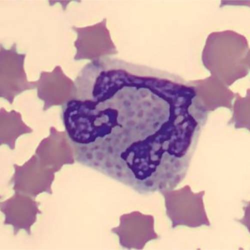
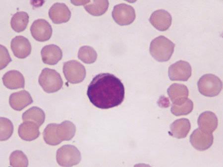
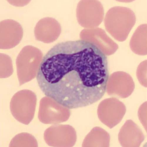
A neutrophil is a polymorphonuclear leukocyte, which means that is has lobulated nucleus — usually 3-5 lobes created by indentations. Neutrophils are inflammatory cells that are often recruited early in acute inflammatory state and play a role in innate immunity, particularly involving bacterial infections. Neutrophils are approximately 10-15µm in diameter and are a useful size marker for assessing the size of red blood cells, which are approximately twice the size of a canine red blood cell.
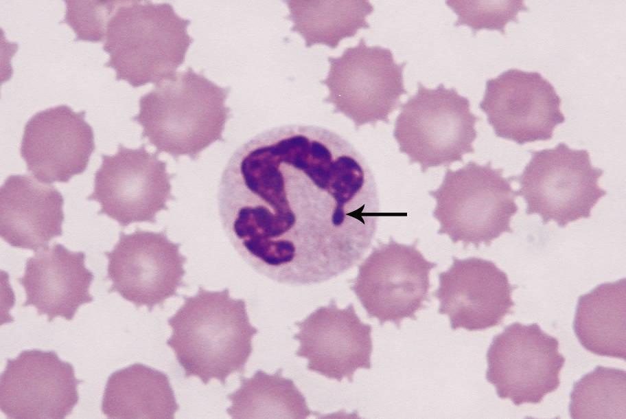
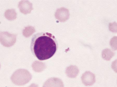
In female dogs and cats, you may see a small number of neutrophils with a small protruberance at one end of the nucleus; this is normal and is the site of the X-chromosome. This feature is called a Barr body (this is illustrated by the arrow in Figure 1).
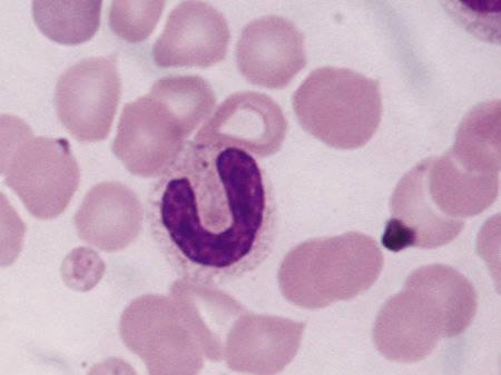
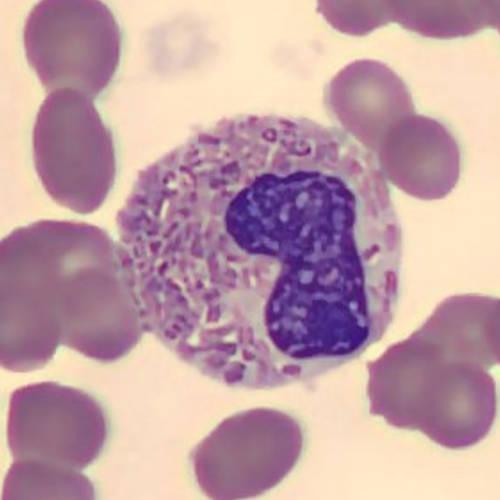
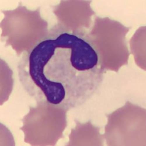
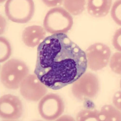
Immature neutrophils are called Band cells and are characterised by an elongated, non-lobulated nucleus that is often "U" shaped, which reaches no more than 50% indentation (Figure 2). A small number of these will be seen in normal animals, and these are new cells that are replacing older ones due to constant turnover of cells.
Eosinophils
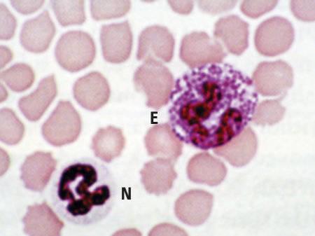
Eosinophils are also polymorphonuclear cells; they are slightly larger than neutrophils, with a segmented nucleus that contains only two or three lobes. They are characterised by numerous, prominent, pink, cytoplasmic granules.
In dogs, the number and size of granules can be variable, sometimes with only a few visible. Greyhounds are unusual in that their granules do not stain at all, so the cytoplasm appears vacuolated. Cats always numerous granules which are rod shaped and are similar sizes (Figure 3).
Remember also that eosinophils are slightly larger than neutrophils, as shown in Figure 4.
Basophils
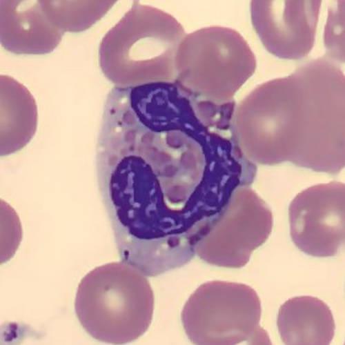
Basophils are the last of the polymorphonuclear leukocytes; they are larger than neutrophils, with long, mildly lobulated, ribbon-like nucleus. They are observed only rarely in blood smears of normal animals, and have dark, purplish cytoplasmic granules against pale grey-blue cytoplasm. The granules are sparse and dark purple in dogs, whereas in cats, they are abundant and pale lilac (Figure 5).
Non-granulocytes
Lymphocytes
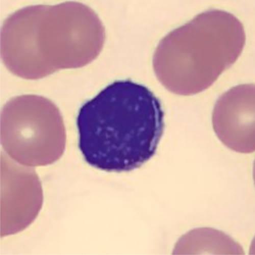
Lymphocytes are a type of mononuclear cell; its nucleus is densely staining, non-lobulated, round to oval shape, with a narrow rim of basophilic cytoplasm (Figure 6).
Small lymphocytes. Lymphocytes vary in size, depending on their activation status. The predominant cell type is a small lymphocyte (slightly larger than a canine red blood cell). These are the most common circulating lymphocyte and are smaller than a neutrophil (Figure 7).
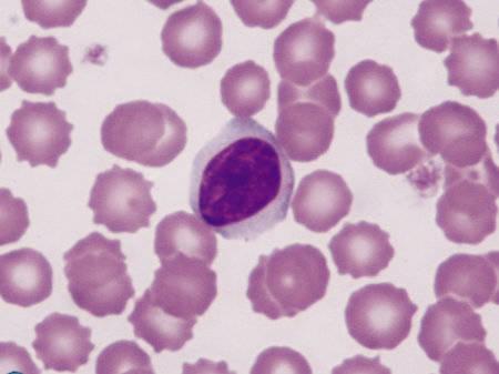
Medium lymphocytes are larger (Figure 8). They have slightly more cytoplasm, which often reaches around the periphery of the nucleus; a small number of these are a normal finding in the circulation and are approximately the same size as a neutrophil.
When lymphocytes are stimulated, then enlarge to approximately 1.5 times the diameter of a red blood cell, these are called reactive lymphocytes and the cytoplasm is more abundant and dark blue (basophilic). These lymphocytes develop due to antigenic stimulation such as an infection (i.e. due to antigenic stimulation), or are seen in neoplastic situations (Figure 9).
Occasionally, large granular lymphocytes are seen; these normally constitute <15% of circulating leukocytes, and function to kill tumour or microbial cells. Prominent cytoplasmic granules are a notable feature (Figure 10).
Monocytes
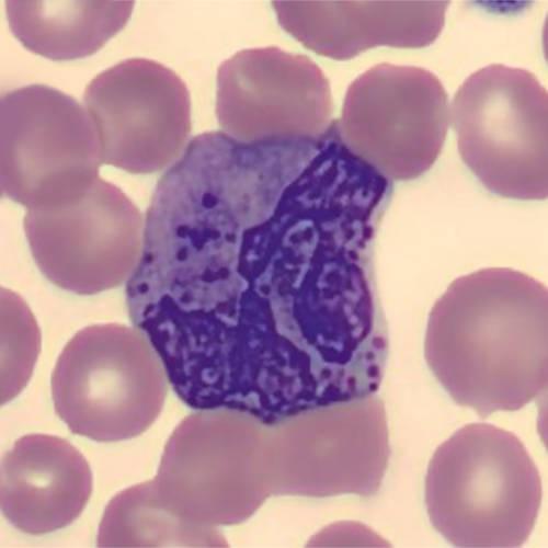
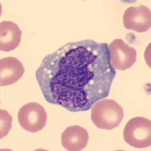
Monocytes are larger than neutrophils with abundant light-blue cytoplasm, sometimes with very small, pinpoint granules. The nucleus shape is variable, such as lobulated, kidney-bean etc. (Figure 11).
Red blood cells (erythrocytes)
Red blood cells (erythrocytes) are the most prominent cell type in the circulation. They function to carry oxgen around the body to ensure adequate tissue perfusion and metabolism.
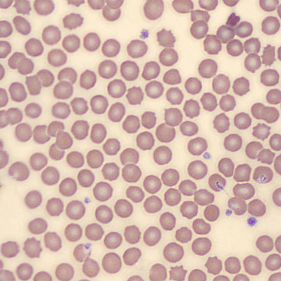
Mature, canine red blood cells (erythrocytes) do not contain a nucleus. They are approximately 7 micrometres in diameter with an area of central pallor (Figure 12).
Feline red blood cells are smaller (5.5 micrometers) and the area of central pallor is less prominent.
An evaluation of red blood cells involves the assessment of:
Firstly, let's reviews some basic terminology for assessing red blood cells:
Variation in cell size. Anisocytosis describes a variation in cell size. Normally, a slight variation will be seen; however, more prominent variations will indicate an abnormality. Anisocytosis is often observed in anaemia. When red blood cells are larger than normal, they are referred to as macrocytic. Conversely, when they are smaller than normal, they are called microcytic (Figure 13).
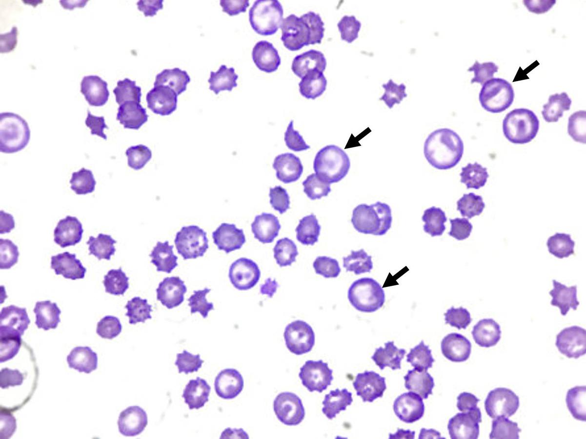
Reticulocytes are immature erythrocytes and a commonly observed macrocytic cell. Normally, less than 1% of all red blood cellls are immature.
Variation in cell shape. A general term for abnormally shaped cells is poikilocytosis. There are a number of different shapes that are useful to recognise, and these will be covered in a following section.
Variation in staining intensity. As well as assessing cell size and shape, the colour of stained cells should be evaluated. For example, reticulocytes stain a blue-grey colour; this is referred to as polychromasia (Figure 14).

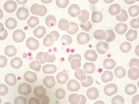
Platelets
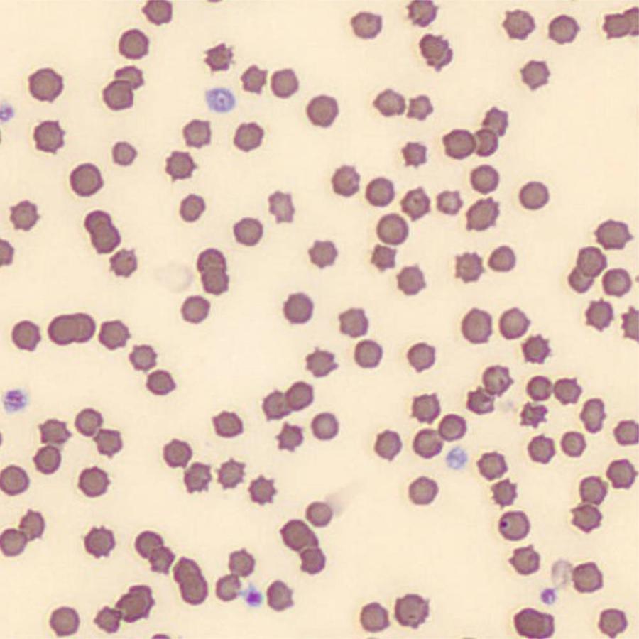
Platelets are small, anucleate, cytoplasmic fragments with clear/pale grey cytoplasm and numerous pink purple granules (Figure 15). They are involved in blood clotting.