

There are a number of common stains that can be used to visualise blood films; which one you use may depend on the availability in your location. The common ones are what's known as Romanowsky stains. Romanowsky-type stains provide good nuclear and cytoplasmic detail. Red blood cells stain red-orange, nuclei stain blue-purple, and cytoplasm stains blue to pink. Commonly-used Romanowsky stains are described below.
Diff-Quik
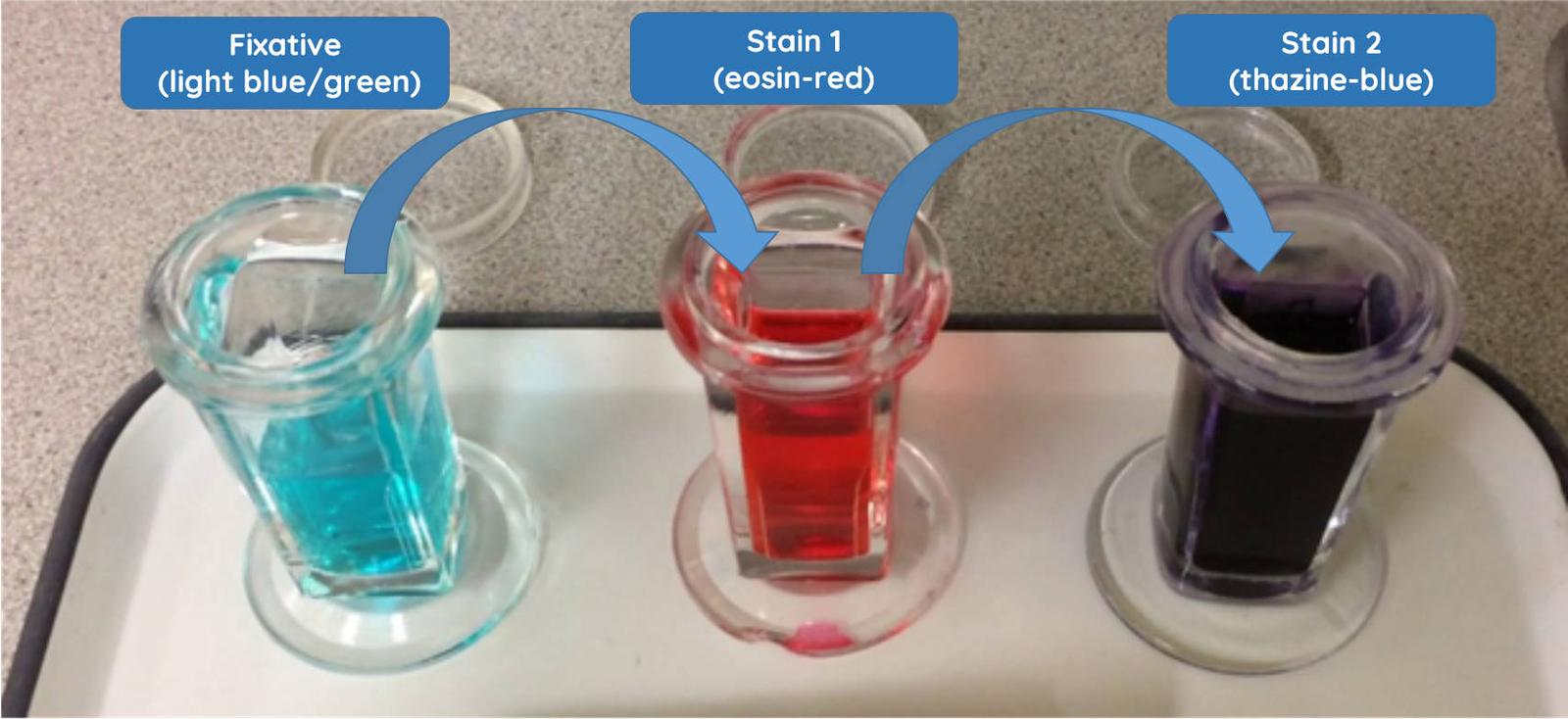
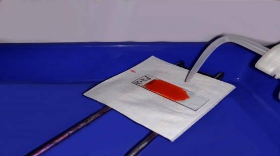
This stain comprises three solutions; a fixative (pale green), Stain 1 (eosin, red) and Stain 2 (thiazine, blue) (Figure 1).
The protocol for this stain is as follows:
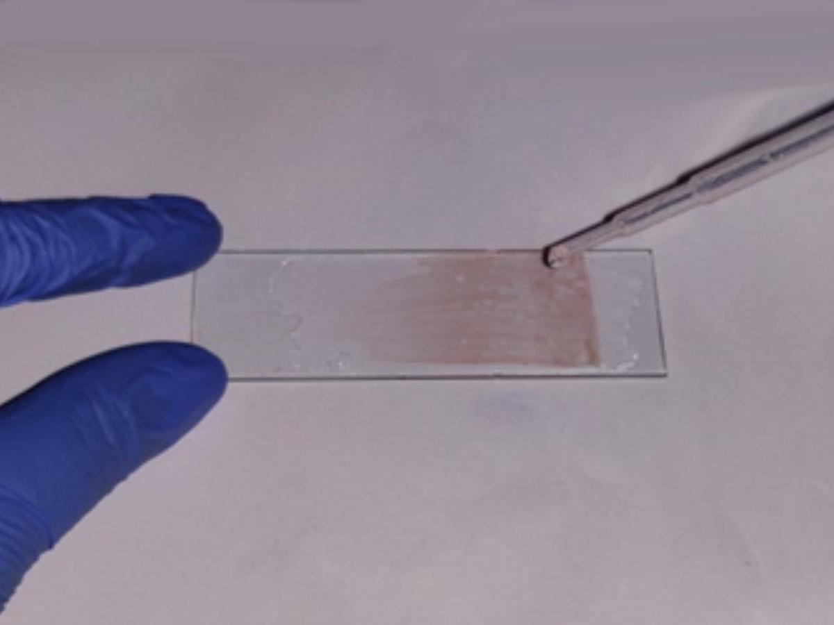
Allow the smear to dry. Always ensure that your blood smear is dry before staining it.
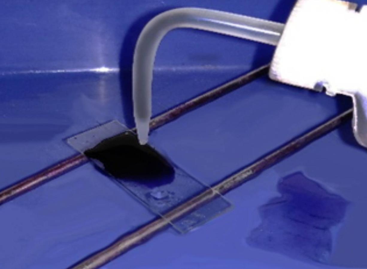
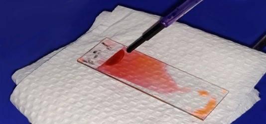
Stain the slide. Dip the slide five times into the fixative (light blue/green) with each dip lasting one second. Allow the excess solution to drain from the slide. Repeat the process for Stain 1 and then again for Stain 2.
Note that it may be necessary to dip the slide seven times in stain 2 in order to visualise platelets sufficiently.
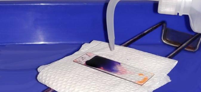
Rinse slide. Once finished, rinse the slide in water, preferably distilled water.
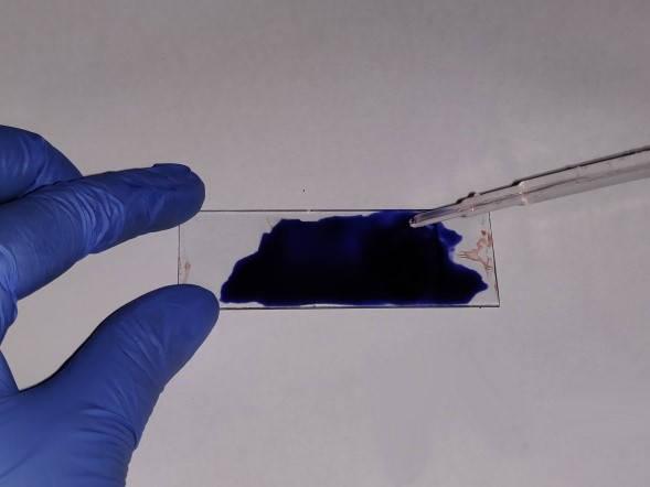
Allow to dry. Allow the slide to air dry in an upright position before examining it.
Giemsa
Giemsa stain can be used for routine examination of a blood film; it is also useful for the identification of haemoparasites.
Allow the smear to dry. Always ensure that your blood smear is dry before staining it.
Methanol fixation. Fix with absolute methanol for one minute by dipping into the solution or dropping the solution onto the slide. Remove the excess solution and then leave to air dry for 30 seconds.
Giemsa stain. Flood the slide with 10% Giemsa stain for 10 minutes (if using 5% Giemsa, leave it for 20 minutes).
Rinse slide. Once finished, rinse the slide in water, preferably distilled water. Take care not to over-rinse, as this can decolourise the slide.
Allow to dry. Allow the slide to air dry in an upright position before examining it.
Field stain
The Field stain is another option for staining blood films; it is often used for detecting malarial parasites in thick blood films.
Allow the smear to dry. Always ensure that your blood smear is dry before staining it.
Methanol fixation. Fix with absolute methanol for one minute by dipping into the solution or dropping the solution onto the slide. Remove the excess solution and then leave to air dry for 30 seconds.
Fields stain B. Cover the slide with Fields stain B (eosin, red colour) for 10 seconds.
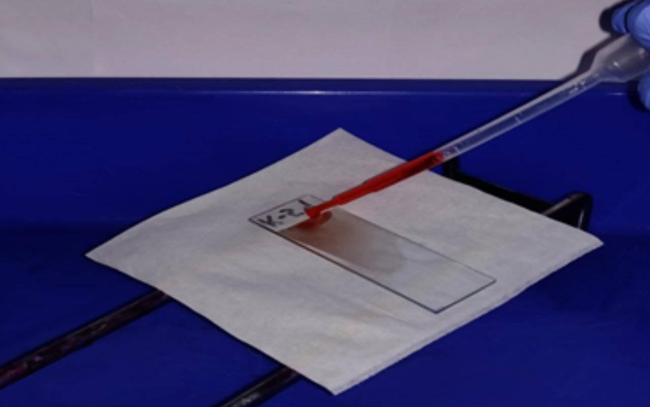
Rinse slide. Once finished, rinse the slide in water, preferably distilled water.
Fields stain A. Cover the slide with Fields stain A (methylene blue, blue colour) for 10 seconds.
Rinse the slide briefly in water (distilled water if possible).
Allow to dry. Allow the slide to air dry in an upright position before examining it.
Leishman's stain
This stain is typically used for staining blood films and for the identification of haemoparasites.
Allow the smear to dry. Always ensure that your blood smear is dry before staining it.
Leishman's stain. Cover the slide with Leishman's stain for one minute; this contains the fixative, methanol. Add double the volume of distilled water to the slide and mix gently by rocking the slide from side to side. Allow the diluted stain to act for 10-20 minutes.
Rinse the slide briefly in water (distilled water if possible). Leave the water on the slide for one minute (the smear should start to turn pink).
Allow the smear to dry. Pour off the water, and allow to air dry in an upright position.