

This last article explains how to access and examine the brain, eyes and spinal cord. Let's start with the brain.
Brain
The brain can be challenging to remove, and proper equipment is needed to do this effectively and safely. Firstly, it needs to be separated from the rest of the body.
Now that the head is removed, the brain can be separated from the skull.
The brain is now removed; the main anatomical features can now be examined.
Eyes
To remove an eye, pick up the lid with rat-tooth forceps (1), and pull gently. With a scalpel, cut carefully into the orbit, cutting the fascia around the eye (2), leaving the eyeball intact (Figure 1).

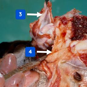
Continue pulling gently on the eyelids (3), and carefully sever the optic nerve at the back of the orbit in the area shown (4), and remove the eye (Figure 2).
Repeat for the other eye.
Spinal cord
The spinal cord is the last tissue to expose and examine.
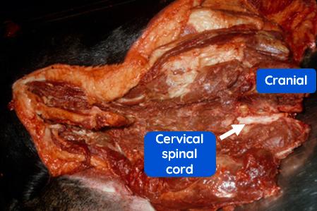
To remove the spinal cord, the commonest method is to remove the skin over the cervical, thoracic and lumbar areas; here the cervical skin is cut away to expose the muscle (Figure 3). The vertebral arches are cut with a de Soutter cutter. (Here the arches have been removed from the atlas and axis vertebrae to expose the cervical spinal cord).
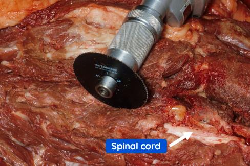
The de Soutter cutter is aligned along the spinal column (in this case the left side) over the vertebral arches, and continued from the cervical to the sacral region. Repeat for the right side (spinal cord, previously exposed).
When the arches have been cut through, the bone forceps are gently inserted into the dorsal spinal canal, being careful not to touch the spinal cord in its meninges. The arches on both sides are severed, and the dorsal spine is retracted.
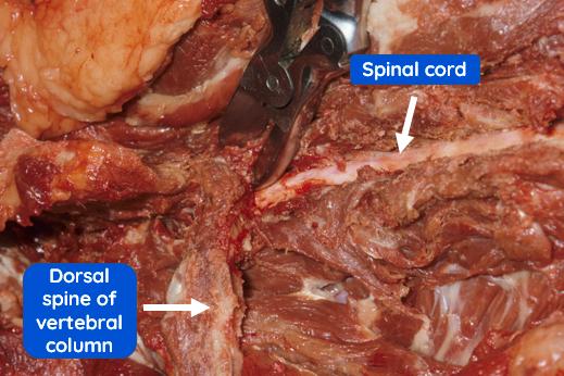
The dorsal part of the vertebral column has been removed to expose the whole length of the cord from the cervical to the sacral region.
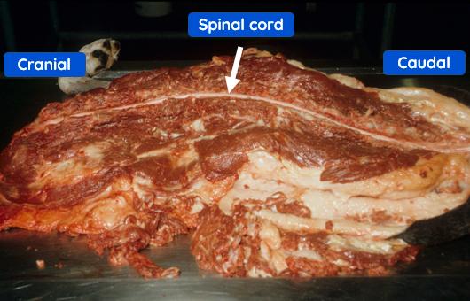
The exposed spinal cord can now be removed
The post-mortem procedure is now complete.