The superficial structures, such as the lymph nodes and salivary glands, will now be examined.
The image below shows the ventral view of the head. First of all, orientate yourself by working out what side of the head is the hand holding.
Submandibular lymph nodes
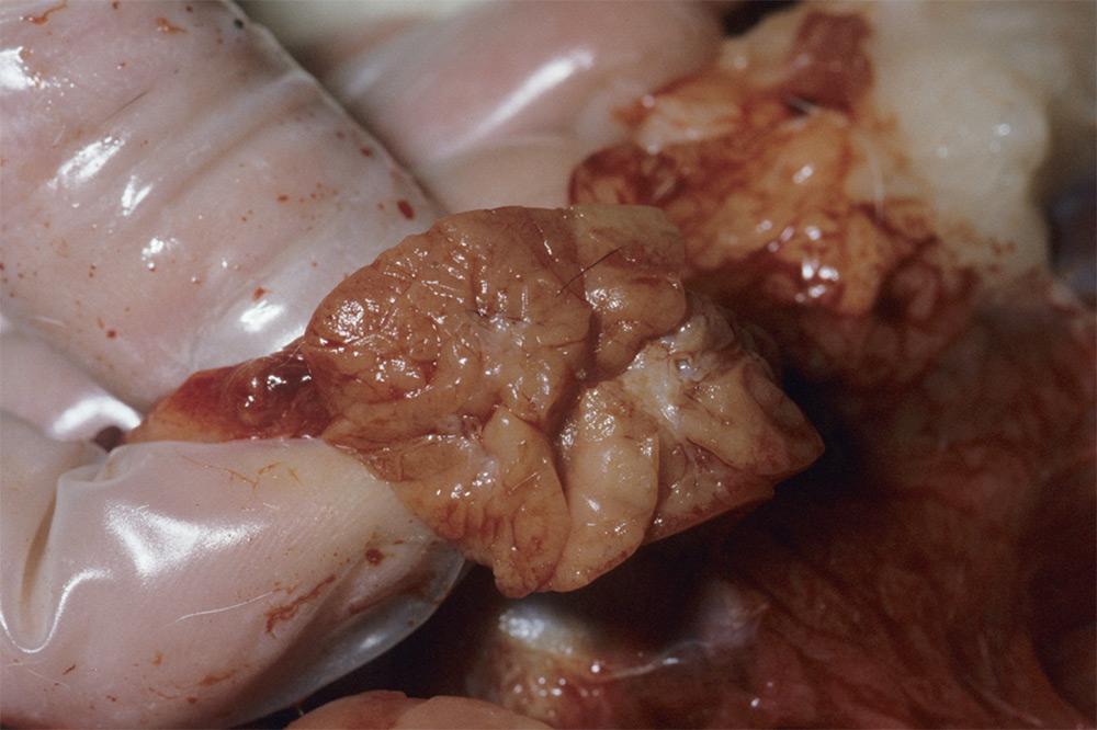
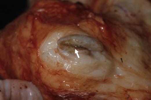
This lobulated pattern is confirmed when the tissue is cut (transected), as shown below.
Axillary and pre-scapular lymph nodes
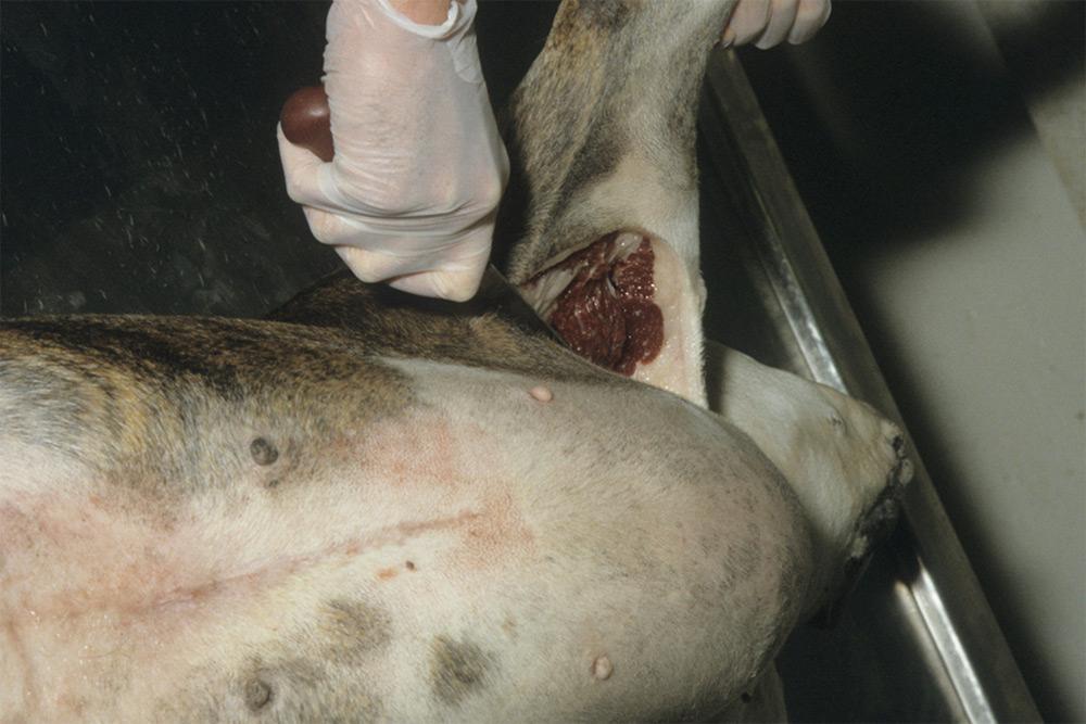
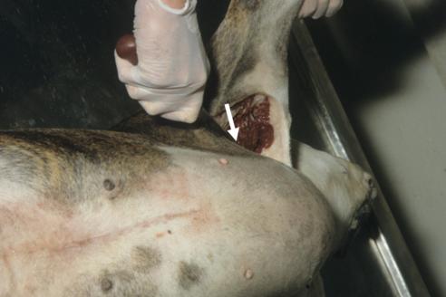
In the image below, the operator is cutting into the right axilla.
The pre-scapular lymph node appears as a pale grey/brown structure in the fat (circle below).
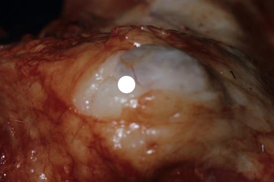
When transected (cut), the cut surface has a mottled grey/brown surface (arrow).
To locate the axillary lymph node, look in the fat and fascia deep in the axilla, in the area shown by the arrow below.
Again, feel for a small, firm nodule (normal size is approximately 1.0 x 0.5 cm in a medium sized dog). It will have the same appearance, when transected, as other lymph nodes.
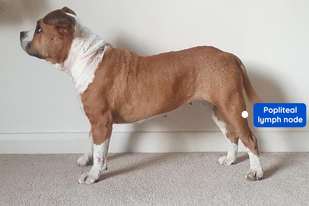
Other superficial lymph nodes
Anther lymph node that should be examined in cats and dogs is the popliteal, which is located caudal and distal to the stifle (see image below).