

Adrenal glands
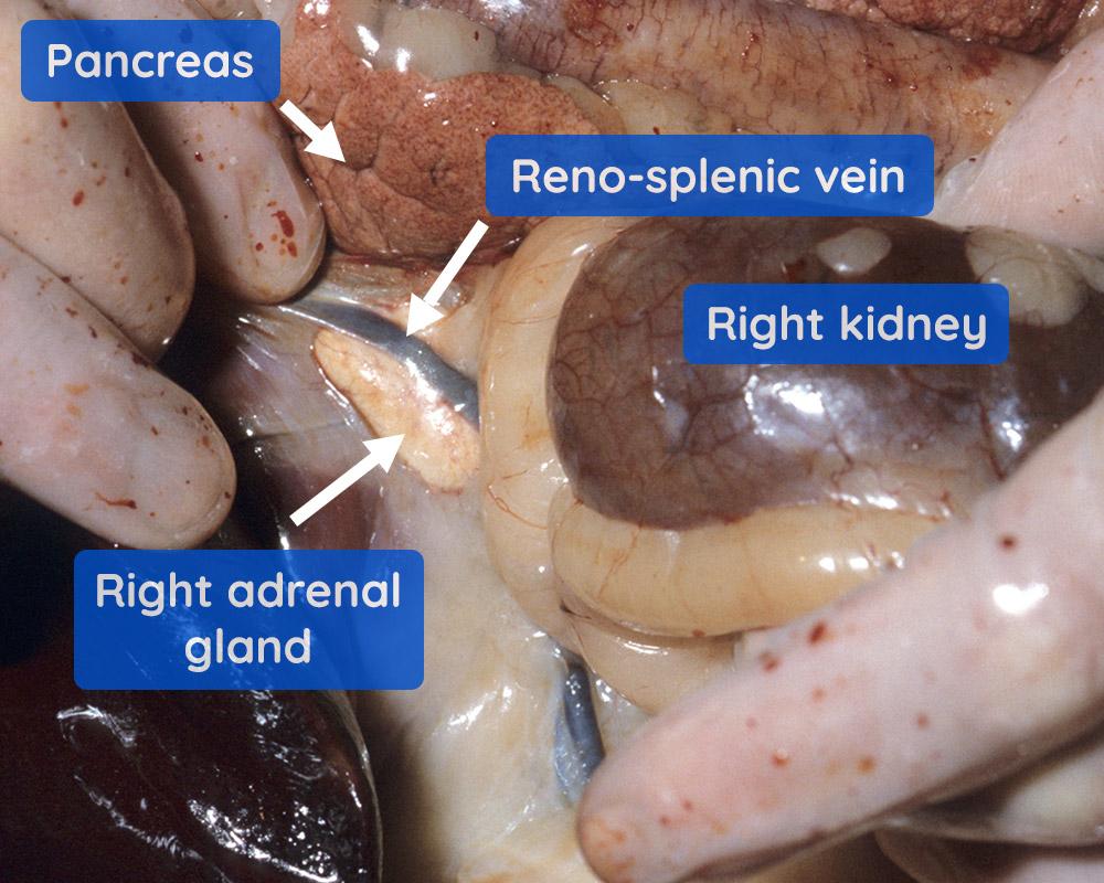
Let's revise where the adrenal glands can be found. They are located at the cranial part of the kidney; the right adrenal gland is shown below (Figure 1).
Once the adrenal glands have been removed, they can be sliced longitudinally and examined:
Urogenital tract
The urogenital tract can be examined partially in situ (bladder, urethra, ovaries and uterus of the female genital tract), and partially following removal of some elements (e.g. kidneys and ureters) from the animal (as we will do in this article). Alternatively, it can be removed intact and then examined.
In this instance, we are removing the kidneys and ureters, leaving the bladder and urethra in situ. The male genital tract is also examined in situ.
Kidneys
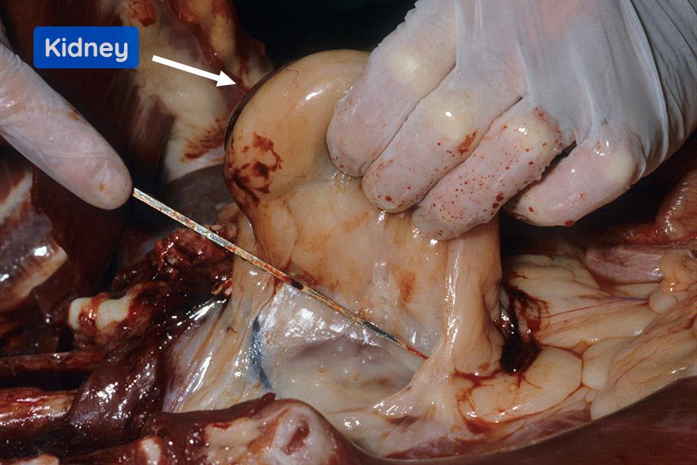
The right kidney is picked up with its surrounding fat. The fat and fascia are then carefully cut away from the dorsal abdomen as shown (Figure 2).
We will explore this area in more detail below:
After the bladder is opened, the urethra is cut open with scissors to the level of the pelvic inlet. In doing this, the prostate gland is sectioned. Examine the mucosal surface of the urethra and the cut surface of the opened prostate.
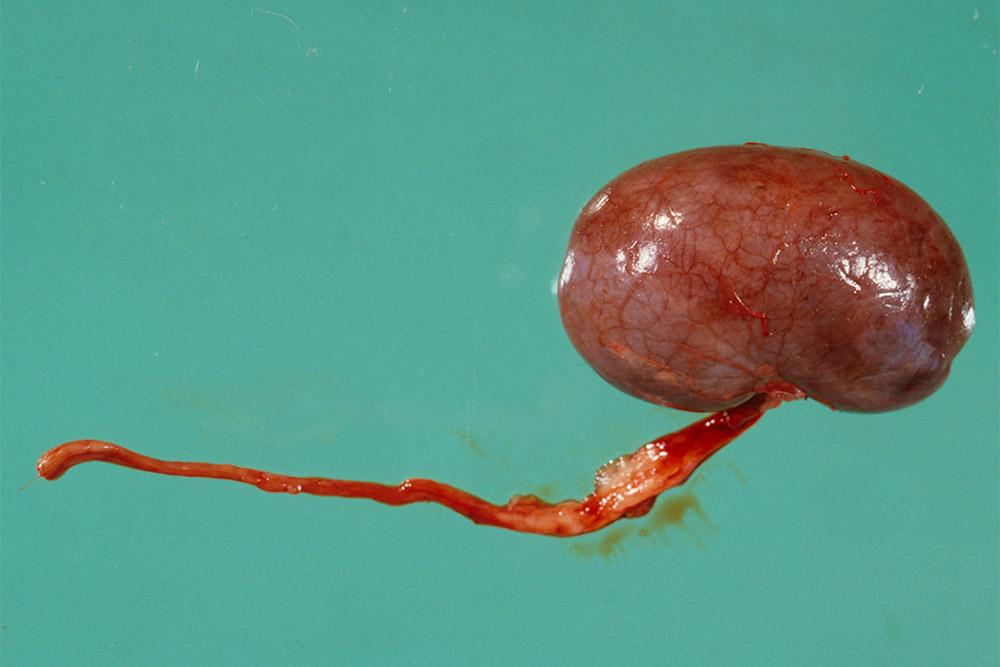
Now examine the removed kidneys and ureters. (Note in this case the fat has been removed) (Figure 1).
The slides below show how to assess the kidneys in detail:
Describe the appearance of this kidney (Figure 4).
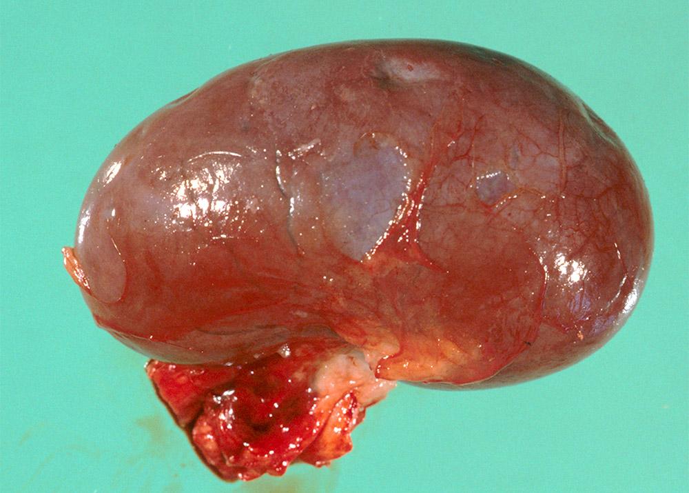
- Measure the dimensions. Using a ruler, measure the length (cranial-caudal), depth (latero-medial), and width (dorso-ventral).
- Describe any gross findings. In this image, the kidney is diffusely brown with a glistening surface and the capsule is easily stripped. There are a few small depressions (approximately 2-3 mm diameter) and a larger, single focus, approximately 5 mm diameter (Figure 5).
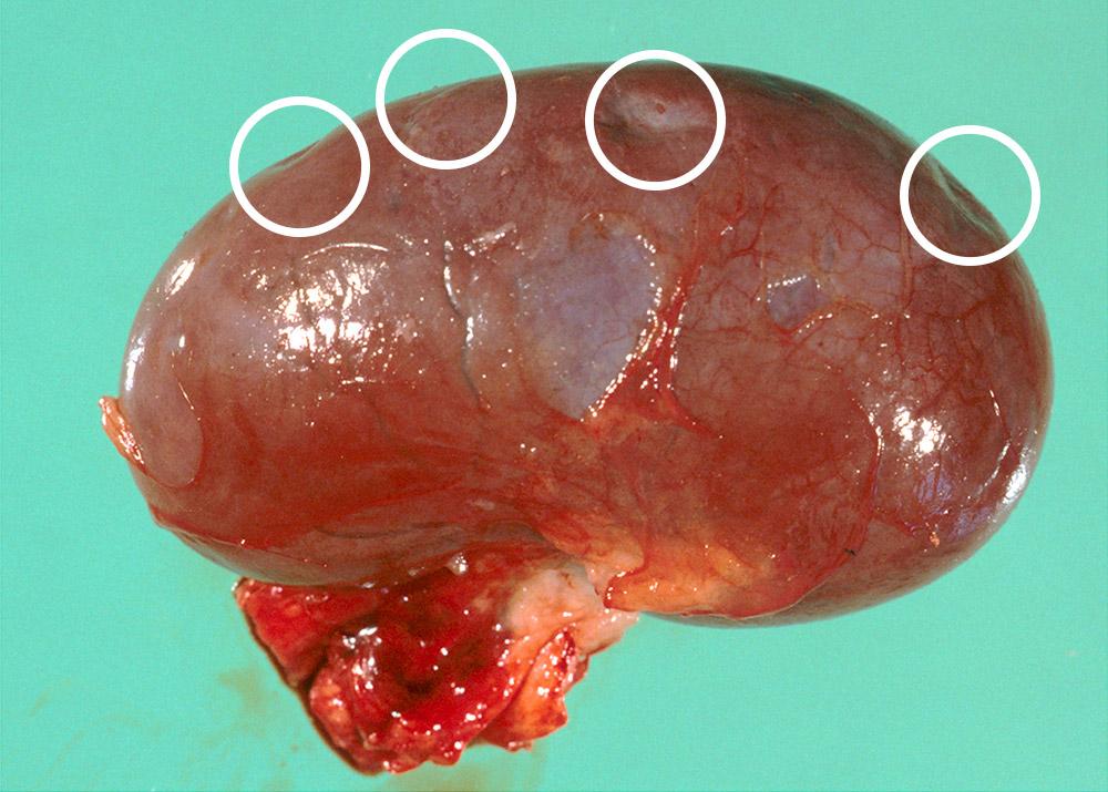
Question: What do these foci represent?
Answer: These foci are depressed, healed scars. They represent earlier infarcts resulting from arterial embolic incidents. They are common in older animals and, in the absence of related clinical signs, represent an incidental finding.
The cut surface of the kidneys can now be assessed:
Iliac lymph nodes
Return to the carcase to identify the external and internal iliac lymph nodes.
The left and right external iliac nodes are situated cranially and laterally to the bifurcation of the aorta, forming the iliac arteries.
The left and right internal iliac nodes are situated posteriorly to the bifurcation of the aorta.
Male genital tract
Now examine the external genital tract, identifying the penile sheath and scrotum . What is the structure indicated by the question mark?
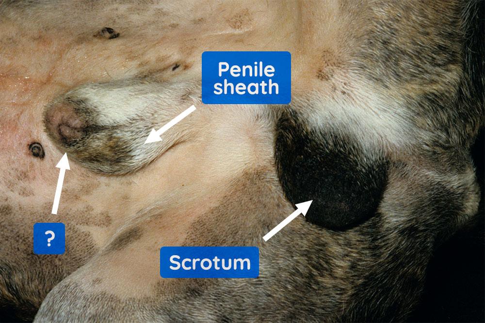
The arrow points to the preputial ring.
Holding the sheath, insert the scissors, carefully, into the prepuce through the preputial ring and cut along its ventral aspect (Figure 7).
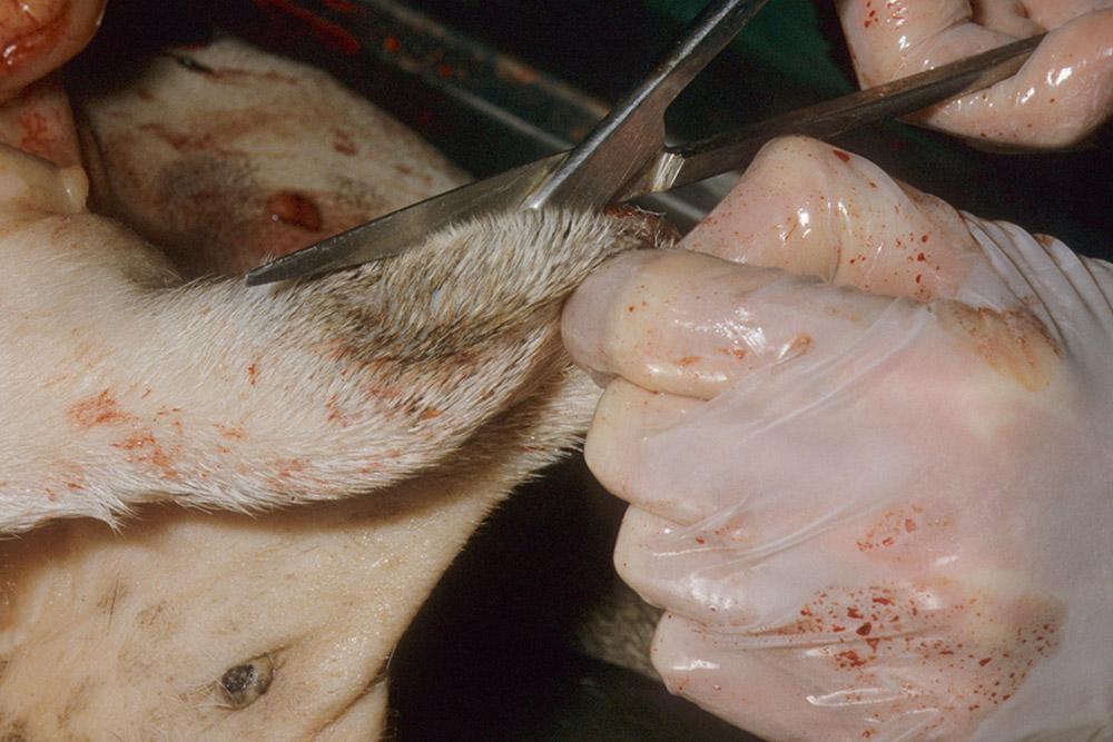
This exposes the penis and preputial surface for external examination.
Insert the scissors into the penile orifice and cut along its length to examine the urethra (not shown).
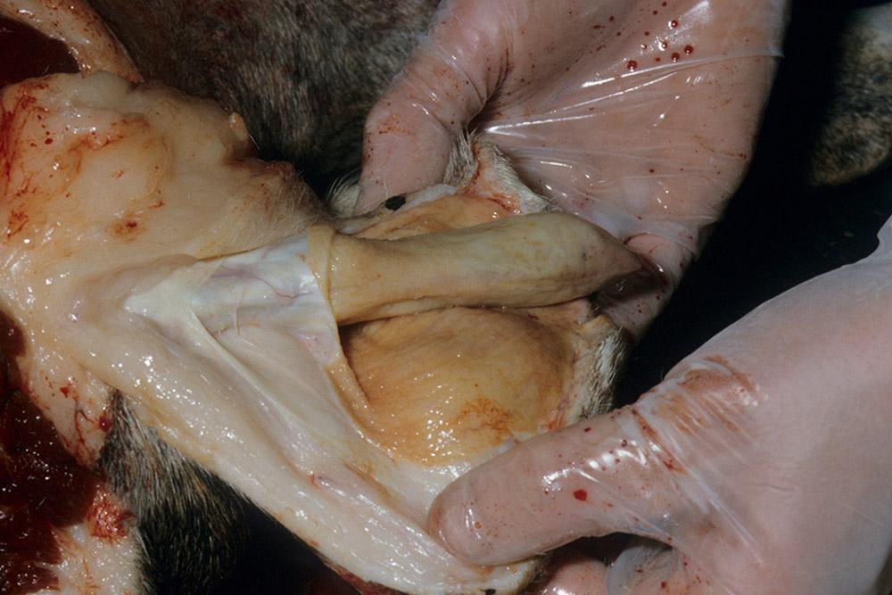
Now let's examine the testes.
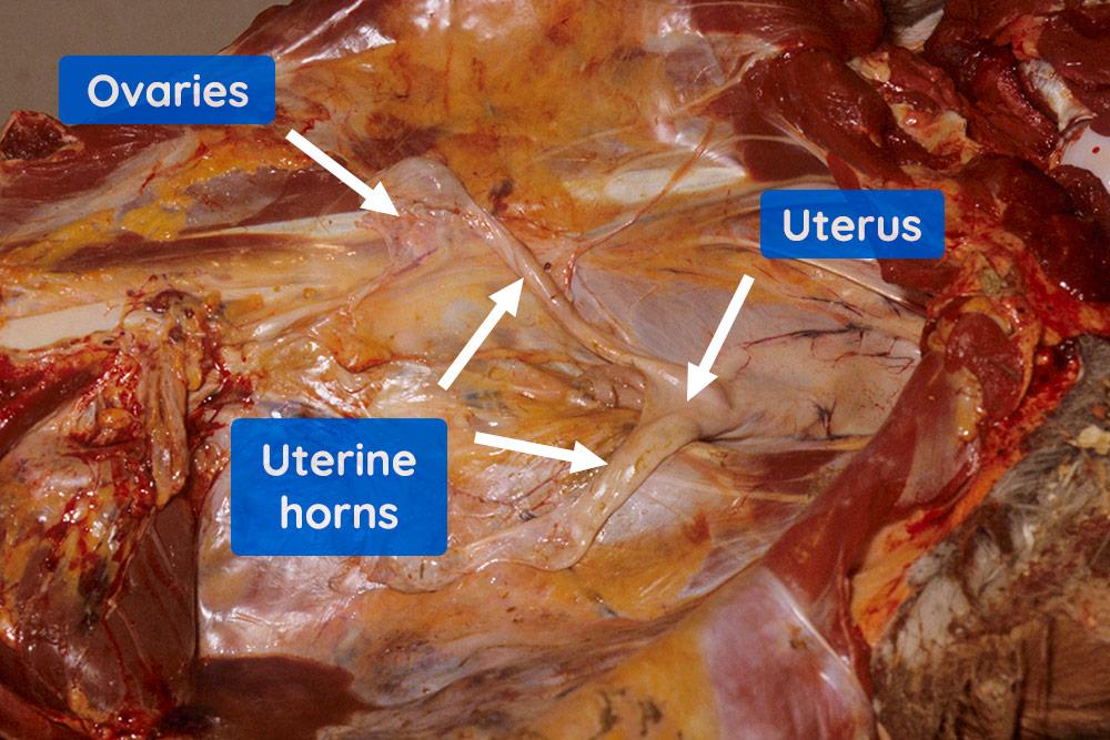
Female genital tract
In a female dog, examine the vagina, uterus, uterine horns and ovaries (Figure 9).
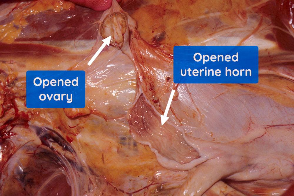
With scissors, cut into and open the uterus (body and horns) (Figure 10) and vagina (not shown); using a scalpel section the ovary.
Removal of the intact urogenital tract
If the urogenital tract (male or female) is to be examined intact (usually because there may be a clinical reason for this), then the following procedure should be followed.
Using small bone cutters, cut through the pelvis. Locate either the right or left acetabular branch of the pubis, cut through into the obturator foramen and continue the cut through the ischial arch. Repeat for the opposite side, being careful not to cut through the urethra and, in the case of the female genital tract, the vagina (Figure 11).
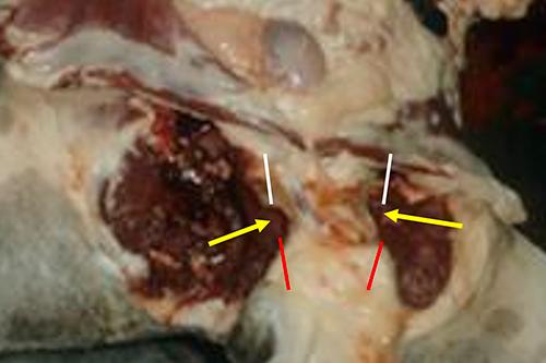
Yellow arrows. Approximate position of the obturator foramina.
White lines. Approximate position of acetabular branches of the pubis.
Red lines. Approximate position of the ischial arches.
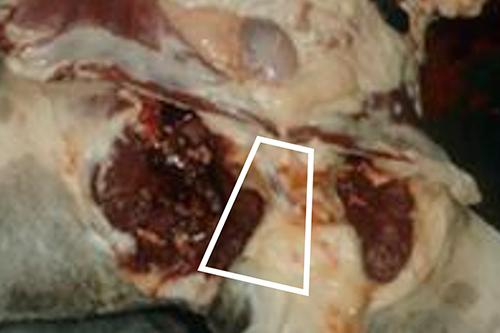
When the cuts are completed, carefully lift the freed piece of pelvic bone (outlined white) with blunt dissection, leaving the urethra/vagina, within the pelvic cavity, intact (Figure 12).
Next, free the kidneys, ureters, bladder (plus uterus and ovaries in the female), and pull them gently away from the pelvis. Cut through the remaining tissues around the anus to free the tract from the carcase. In a male dog, free the penis and trace the subcutaneous urethra to the level of anus. The entire tract can then be carefully cut free from the carcase, around the anus. It is then examined intact, following similar procedures to those described above, for the in situ examination.