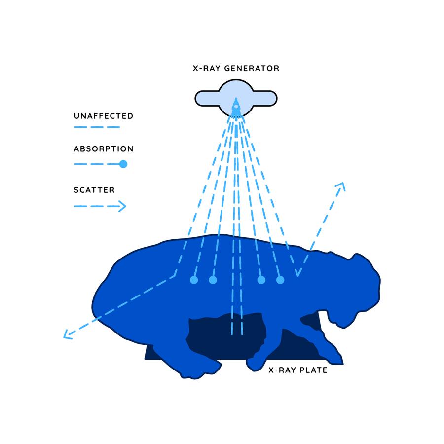
When the beam is produced by the generator, it passes down through the patient and is altered by interacting with tissues in a number of ways. Energy is lost from the beam by these interactions. The remaining x-rays hit the detector plate, creating the image.
Absorption. As the primary beam passes through the patient, some energy is absorbed by tissues and thus removed from the beam. Denser tissues, such as bone absorb more energy than less dense structures like muscle or fat. As a result, where an x-ray passes through bone, less gets through to the detector.
Scatter. Some energy in the beam may bounce off the patient as scatter radiation. This energy is a hazard to any staff in close proximity to the x-ray machine, and thus good shielding and PPE are needed. The higher the kV setting, the more scatter radiation will occur, and the image quality will also deteriorate, with reduced contrast.
Intensifying screens. Where the depth of the patient is over 10cm, we can use an intensifying screen when performing an x-ray, which increase the energy produced by photons in the beam and reduce scatter of radiation. This allows us to reduce the dose of radiation and shorten the exposure time, resulting in less motion blur and less scatter. These screens do cause some resolution loss though.