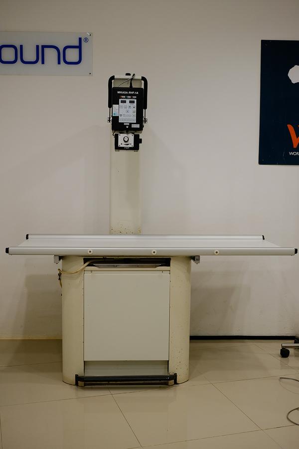
Generating the beam
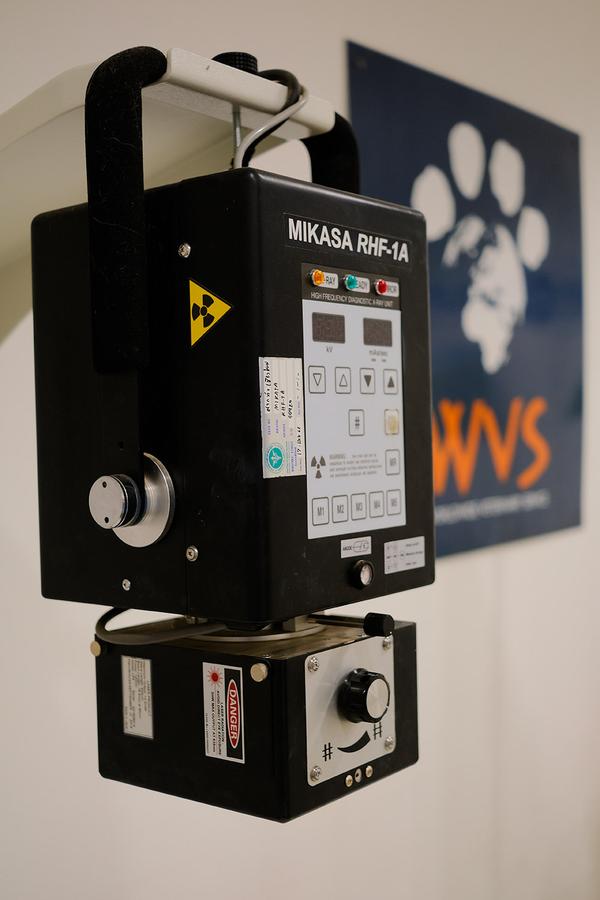
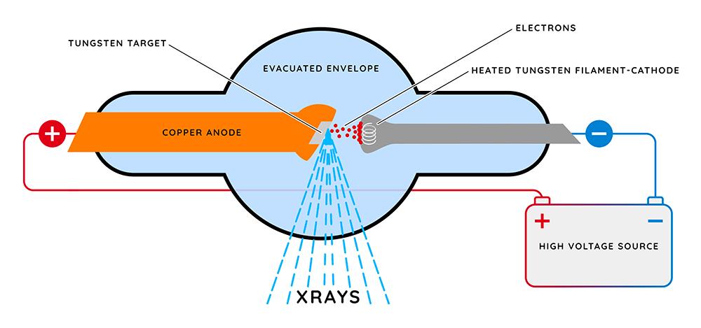
The x-ray beam is produced by a generator within the x-ray machine. When the x-ray button is pressed, an electric current in the form of electrons passes through the circuit within the generator, hitting a tungsten target, which results in the release of photons (tiny particles of radiation). These travel out of the generator in a straight line (usually downwards towards the floor) and make up the primary x-ray beam.
By manipulating the current and voltage settings on the x-ray machine, the clinician alters the properties of the primary beam produced. As a result, different settings produce different primary beams (more or fewer photons being released over a period of time), which allows the clinician to adjust the beam to produce the best image for a certain part of the body. This is because different organs have different densities, and thus need a greater or lower amount of penetration by the beam to pass through the patient. A simple example for this would be that a stronger x-ray beam would be needed to penetrate through a dense substance such as bone (which absorbs lots of the beam), than a less dense substance such as soft tissue (liver, muscle etc).
Film focal distance
The detector is placed directly under the generator (where the primary beam exits), and the patient is placed on top of the detector. The film focal distance is the distance between the focal point on the generator and the detector plate underneath the patient. This ensures the radiographs produced are in focus. The actual distance varies between generators and is usually set when the x-ray machine is installed.