

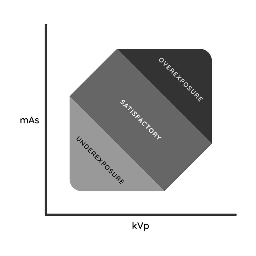
To manipulate the beam and alter the final image, we can change three exposure settings on the X-ray machine: kV, mA, and seconds. In practice mA and seconds are typically combined as a single value, mA/seconds (mA/s), and so the practitioner adjusts the kV and mA/s for each radiograph.
kV (Kilovolts)
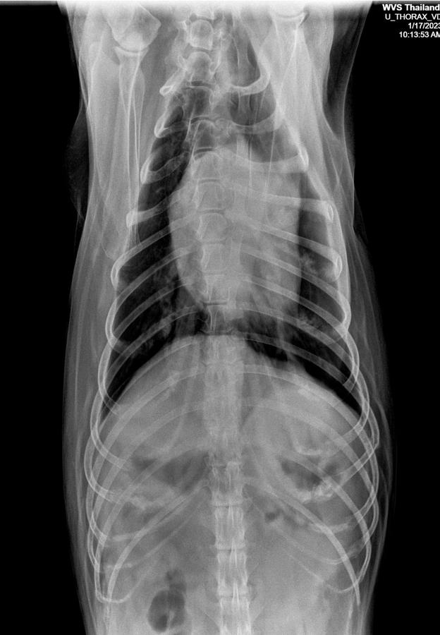
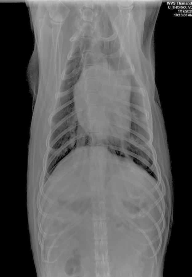
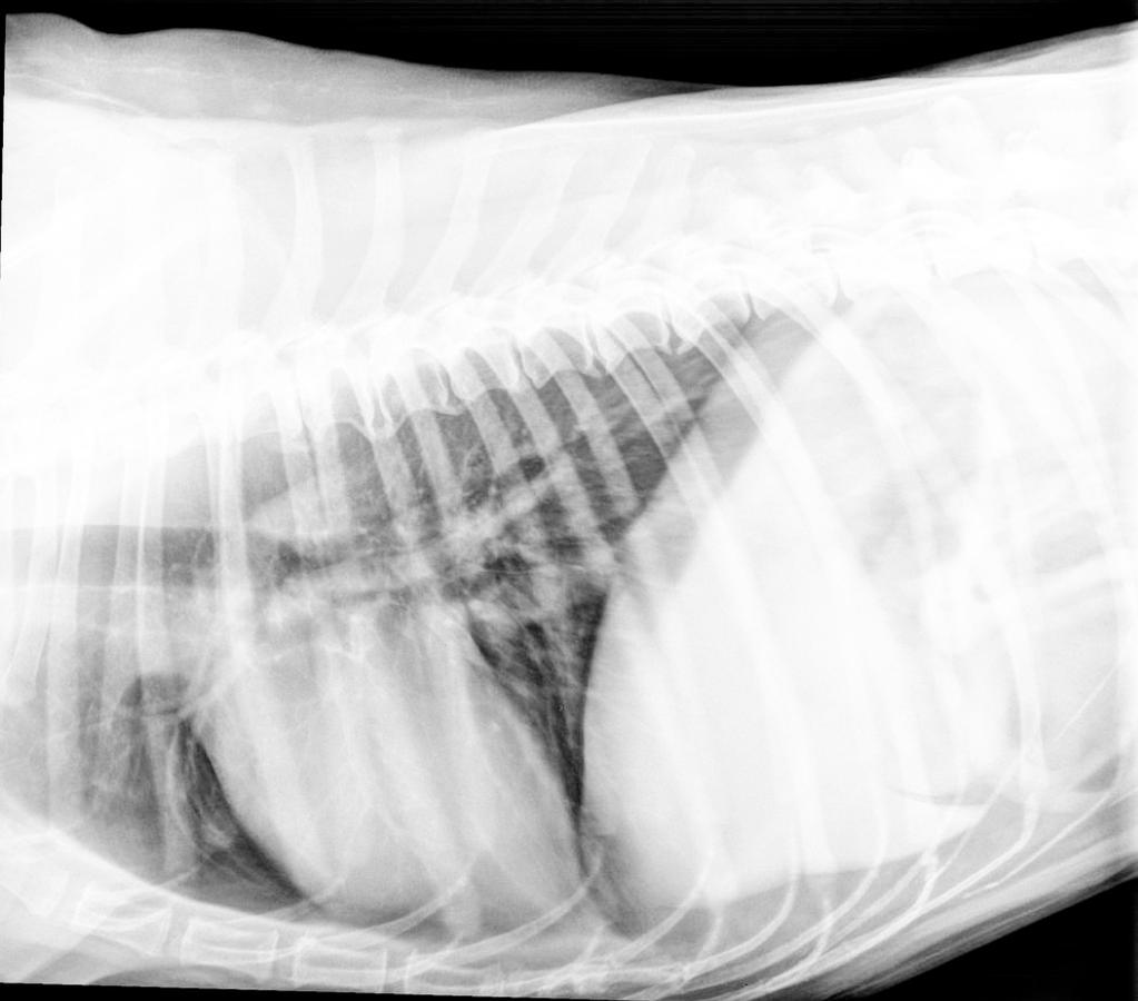
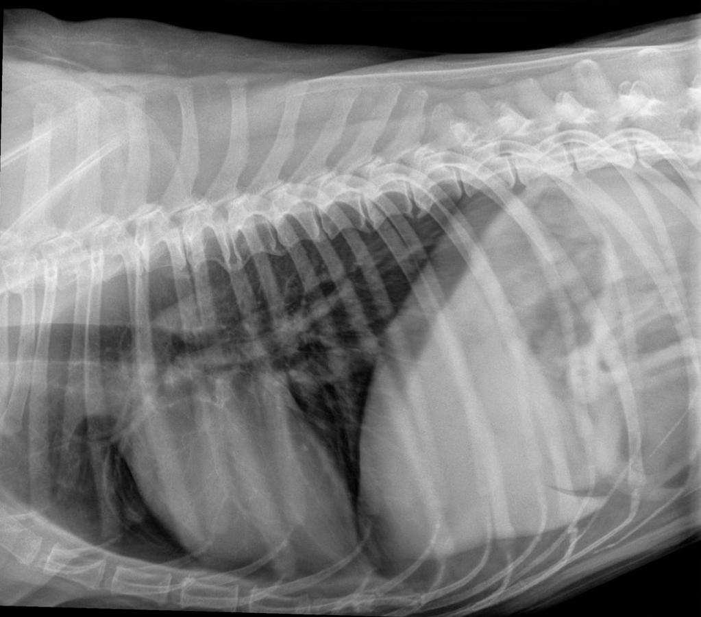
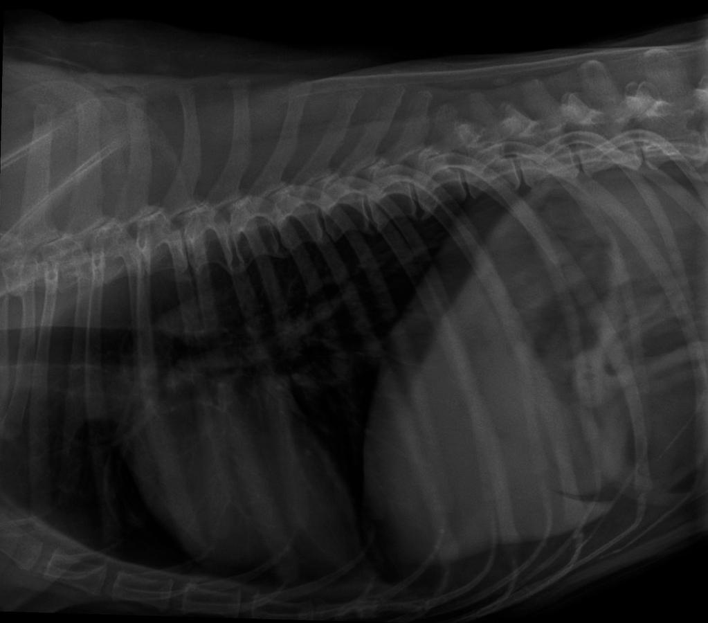
This is the voltage applied, which changes the strength of the x-ray primary beam produced. Changing the kV affects the quality of the x-rays. A higher kV setting increases the energy of the photons released, meaning a stronger and more penetrating x-ray beam is produced. A higher kV setting is used when imaging larger animals (to ensure the beam penetrates all the way through the animal), and when imaging parts of the body with varied tissue densities. For example, the thorax has bone, soft tissue and gas, and so requires a higher kV than the abdomen which is mainly soft tissue and a similar density throughout. Increasing the kV does, however, reduce contrast in the final image. This means it becomes more difficult to distinguish between areas of interest and the rest of the image, and so this may reduce the diagnostic quality. As a result, a compromise must be reached. The kV must be high enough for adequate penetration, but not so high as to lose contrast.
mA (Milliamperes)
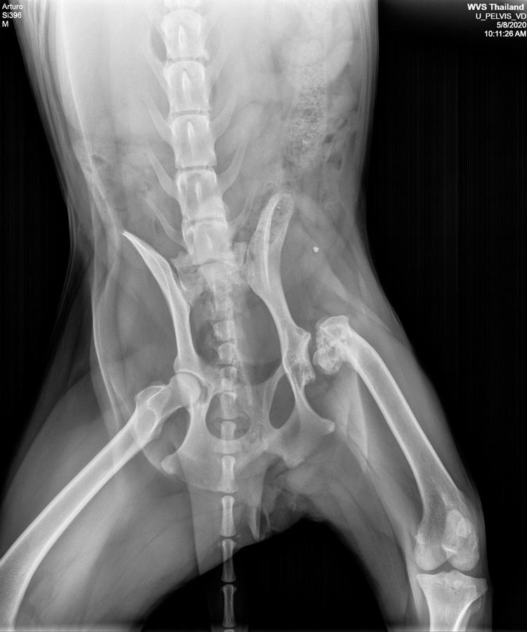
This is the current applied within the generator. An increase in mA increases the number of x-ray photons produced, without affecting the energy spectrum of the individual x-rays. This increase leads to increased image contrast, which is desirable as it improves the ability to distinguish between areas of interest in the radiograph, and thus improves the image's diagnostic quality. This increase in contrast is most useful when imaging areas of the body with roughly similar densities. For example when radiographing the abdomen, which is made up of lots of soft tissue elements (liver, stomach, spleen), it can be difficult to tell these apart from one another. By increasing the mA setting this becomes more achievable. However, an increase in the mA also increases the heat produced in the generator. This limits exposure time and reduces the generator tube's life span. It also means that the patient is exposed to a greater amount of ionising radiation, and so this should be set as low as reasonably practicable.
Exposure time (seconds)
This affects how long the x-rays are being produced for. The longer the exposure, the more photons are released and the more radiation hits the patient and thus the plate underneath. The variable mAs (milliampere-seconds) is the product of exposure time and milliamperes. The shorter the exposure time, the lower the chance of any movement blur occurring in the photo, but the higher the mA must be to achieve the same level of contrast. The longer the exposure time, the more likely motion blur is to occur, but the mA may be reduced which is safer for the patient. For taking radiographs of highly mobile parts of the body (such as the thorax), a short exposure time is particularly useful to reduce motion blur, whereas for parts of the body that are less rapidly moving (abdomen), the practitioner may use a longer exposure time.

Practitioners commonly use a technique chart, with standardised exposure settings that are applied when taking a radiograph. These settings are unique to each x-ray machine, and based on the species of animal, the site radiographed and the radiographic view (explained later). We have added two downloadable exposure charts below, with suggested kV and mA settings for both 80cm and 100cm focal distance X-ray devices, with an explanation of how to adjust the settings if using a grid. These may be used as a rough starting point to develop your own exposure chart, due to the individual differences between each machine.