

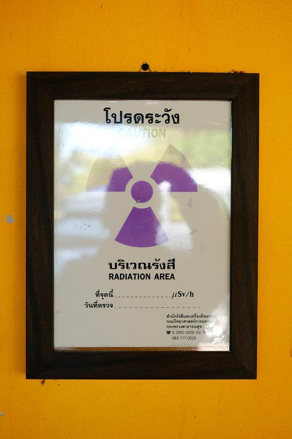
Radiation
Ionising radiation in X-rays interacts with body tissues, damaging and changing the DNA within cells. At a low level, the risk of these changes causing harm is slim, however, frequent X-ray exposure to humans or animals can lead to negative health impacts in the long term. This includes an increased risk of cancer, even years down the line.
Practitioners who regularly produce radiographs for diagnostic purposes are most at risk of receiving low-level scatter radiation. This exposure to scatter radiation may be repeated over time resulting in high levels of harmful radiation. For this reason, practitioners follow a number of safety principles to minimise radiation exposure, and thus reduce the risks to their long-term health.
The aim is to keep radiation exposure as low as reasonably achievable (ALARA).
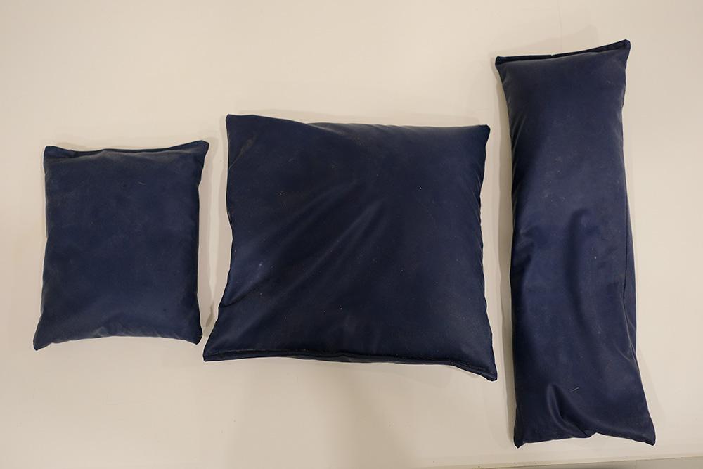
Avoid the primary X-ray beam
The practitioner should never place any of their body within the primary beam, which carries the highest dose of radiation. Positioning aids such as sandbags and wedges can be used to keep patients in the correct positions, eliminating the need for practitioners to be within the primary beam. Always collimate appropriately so that the primary beam is as small as possible whilst remaining diagnostic.
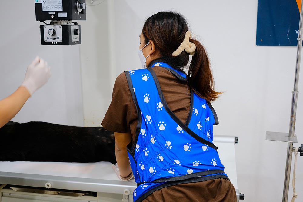
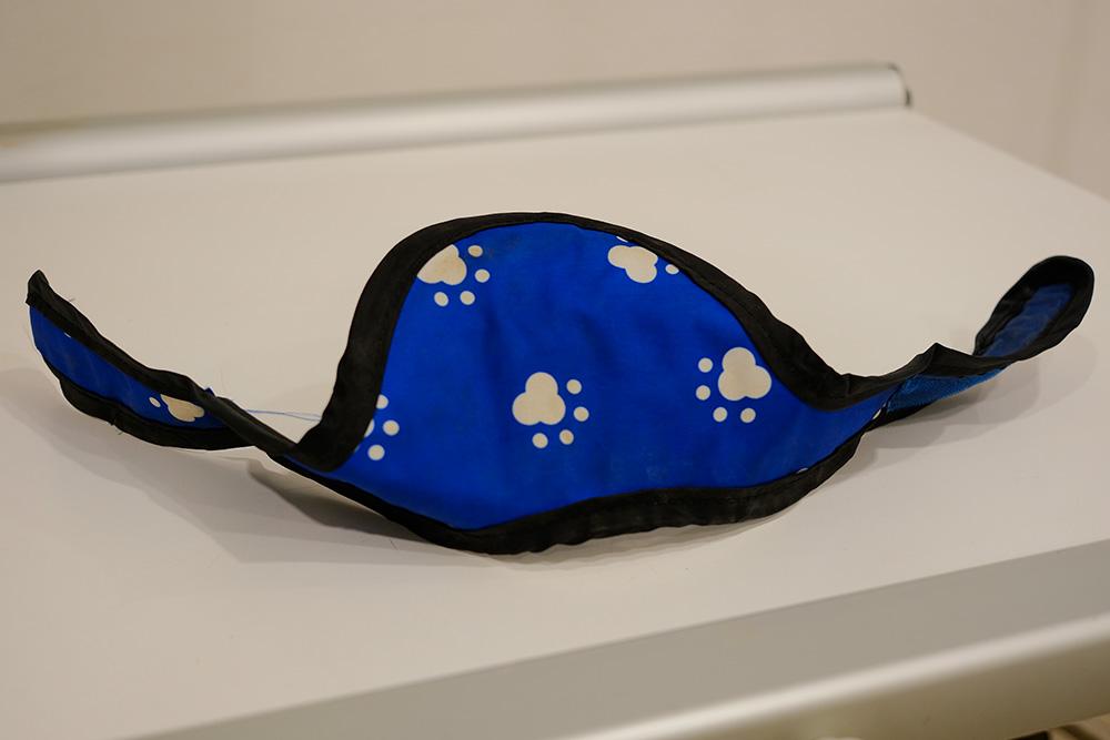
Use shielding and wear personal protective equipment
High-density metals, such as lead, are effective at blocking radiation exposure. Where possible, the practitioner should stand behind a radiation shield which contains at least 0.5cm of lead. Practitioners may also wear equipment including a lead apron, lead gloves and a thyroid protector. A lead matt placed beneath the X-ray plate also helps reducing scatter radiation. Practitioners should always avoid the primary beam as some radiation may still get through protective equipment. In some circumstances, practitioners may hold conscious patients, but make sure they wear appropriate shielding and always stay out of the primary beam.
Increase distance from X-ray generator and use shielding
Doubling the practitioner’s distance from the source of radiation decreases its intensity by 4 times (inverse square law). This means the practitioner should always maximize their distance from the source to minimise exposure.
Ensure X-rays are justified
For example, only use radiography where the suspected differential diagnoses may show up on a radiograph (e.g. Gastric Dilatation and Volvulus). In the case of free abdominal fluid, ultrasound is more specific and is usually more likely to give a diagnosis than a radiograph, without carrying the risk of radiation (and sedation).
Reduce time spent exposed to X-rays
This may be done by having multiple operators who perform X-rays at different points in time. Taking note of which staff were present at the time of exposures, whilst also recording exposure settings used lets us monitor which staff members have been exposed to more radiation. Dosimeters worn by all members of staff involved in taking radiographs are a good way of keeping track of radiation exposure. If dosimeters quickly fill with radiation, it suggests an issue in safety protocols within the practice.